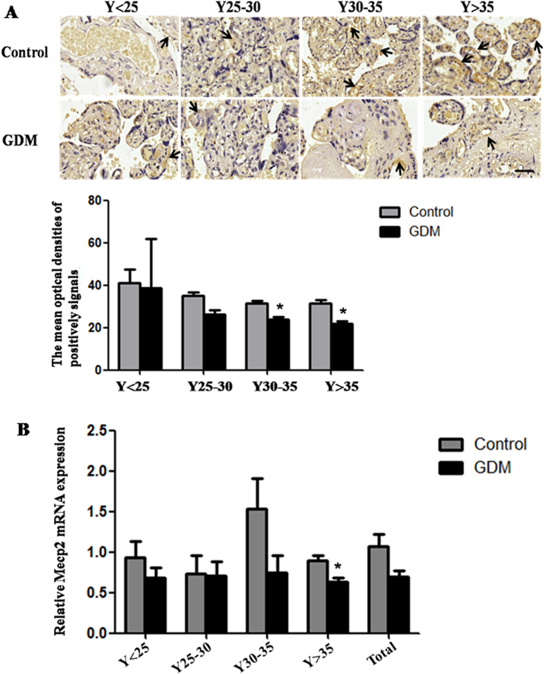
Figure 6. The expression of Mecp2 in the placental tissues from patients with GDM.
(A) The protein level of MECP2 in the placental tissues from patients with GDM and normal pregnant women was detected by immunohistochemistry. The stain was developed with DAB and cell nuclei were stained with haematoxylin. Black arrow indicated positive signals and the positive signals of MECP2 are brown. The histogram represents the MODs of positive signals of MECP2 in placentas. Scale bar = 50 μm. (B) qRT-PCR was used to detected the mRNA level of Mecp2 in the placental tissues from patients with GDM and normal pregnant women. Gapdh serves as an internal reference. *P < 0.05.

