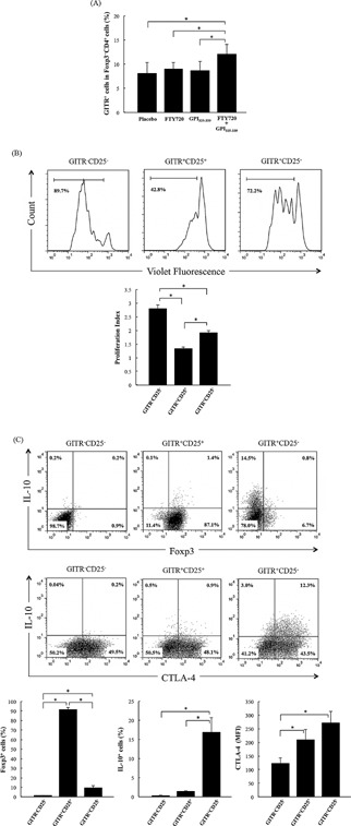
Figure 5.

Combination treatment with FTY720 plus GPI325‐339 induces GITR+ non‐Treg cell populations in inguinal lymph nodes. Cells were obtained from inguinal lymph nodes of treated mice at day 14 (completion of treatment). (A) The percentage of GITR+ cells in Foxp3−CD4+ cells was determined by flow‐cytometric analysis (four animals from one experiment). (B) Violet Fluorescence‐labeled GITR−CD25−CD4+ cells (5 × 10⁴ cells) were cultured with unlabeled‐GITR−CD25−CD4+ cells, GITR+CD25+CD4+ cells, or GITR+CD25−CD4+ cells (2.5 × 10⁴ cells) in the presence of IA/IE+ cells (5 × 10⁴ cells) and anti‐CD3 mAb (1.0 μg/mL) for 72 h. Proliferation index was analyzed by FlowJo software (five animals from one experiment). (C) GITR−CD25−CD4+, GITR+CD25+CD4+ cells, and GITR+CD25−CD4+ were stimulated with anti‐CD3/anti‐CD28 coated dynabeads and IL‐2 (50 U/mL) for 72 h. The percentage of Foxp3+ cells and IL‐10+ cells and mean fluorescence intensity of CTLA‐4 expression were determined by flow‐cytometric analysis (three determinations). The results are shown as mean + SD. The significance of differences was examined by Duncan's test (*denotes P < 0.05).
