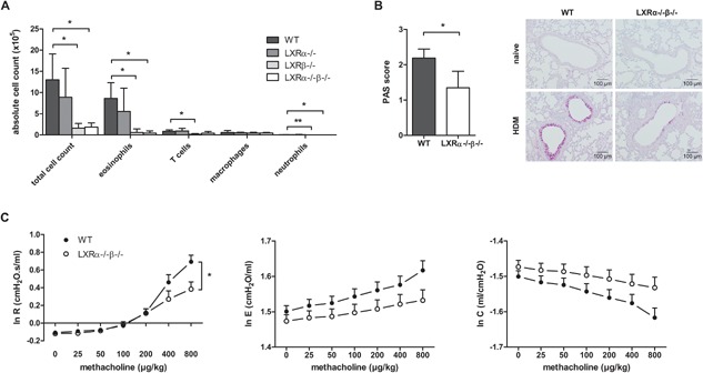
Figure 2.

LXR‐deficient mice display reduced airway inflammation, goblet cell hyperplasia, and airway hyper‐reactivity (AHR) in house dust mite (HDM)‐induced asthma as compared to wild‐type (WT) mice. WT, LXRα−/−, LXRβ−/−, and LXRα−/−β−/− mice were sensitized against HDM and subsequently challenged intranasally with HDM for 4 consecutive days. (A) Differential cell counts in the bronchoalveolar lavage measured by flow cytometry. (B) Mucus production evaluated by PAS staining of lung paraffin sections. (C) Dynamic airway resistance [R], elastance [E], and compliance [C] was measured in mice exposed to increasing doses of methacholine after HDM challenge. (A) A Kruskal–Wallis test followed by a post‐hoc Dunn's test was used. (B) Statistical analysis between two groups was performed using a Mann–Whitney U test. Data are shown as mean ± SD, n = 5–7 mice per group. The results show one representative experiment out of at least two independent experiments. (C) The repeated measures in dynamic AHR of one independent experiment were analyzed by residual maximum likelihood. Data are shown as mean ± SE, n = 5–7 mice per group. Significant P‐values were ranked as P < 0.05 (*) and P < 0.01 (**).
