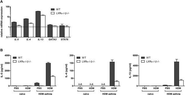
Figure 4.

LXR‐deficient mice show decreased Th2 cytokine expression in the lungs and lung‐draining lymph nodes as compared to wild‐type (WT) mice in OVA‐induced and house dust mite (HDM)‐induced asthma. (A) mRNA expression levels of Th2‐associated cytokines (Il‐5, Il‐13, Il‐4) and transcription factors (Gata3 and Stat6) in the CD4+ cells of the lungs of WT and LXRα−/−β−/− mice after two consecutive OVA aerosols. Lung cells were pooled per group before CD4+ enrichment and relative mRNA expression levels were determined in triplicate by qPCR. (B) Cytokine production by mediastinal LN cells of WT and LXRα−/−β−/− mice after four intranasal HDM challenges. Mediastinal LN cells were pooled per group and restimulated in quadruplicate with HDM for 48 h. Cytokine levels were measured by luminex analysis. Data are shown as mean ± SD, n = 3–7 mice per group. The results show one representative experiment out of two independent experiments.
