Figure 3.

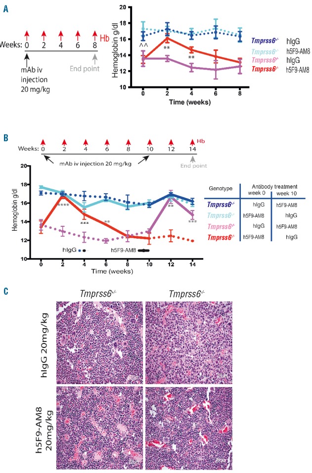
Anti-HJV antibody increases hemoglobin and recovers erythropoiesis in a non-inflammatory high hepcidin model. (A) Schematic summarizing experimental protocol (n=10). At 0 weeks hemoglobin levels of the homozygous Tmprss6−/− was significantly lower than the heterozygous Tmprss6+/−. At week 2–6 the Tmprss6−/− mice had a significant increase in hemoglobin levels in comparison to the Tmprss6−/− hIgG mice. (B) Cross-over experiment (n=5). Homozygous Tmprss6−/−mice treated first by the humanized istotype control mAb hIgG were treated at week 10 with 20 mg/kg iv h5F9-AM8. Anemia was corrected again in weeks 12 and 14. Change from baseline data over time were analyzed using mixed model (fitting change from baseline by timepoint and using baseline as a covariate), **P<0.01, ***P<0.001, ****P<0.0001. Statistical analysis shown is for comparisons between Tmprss6−/− mice treated with hIgG and h5F9-AM8. Statistical analysis for comparisons between Tmprss6+/– and Tmprss6−/− mice treated with hIgG at time 0 is shown by ^^, P<0.01. Data in A and B are presented as mean±SEM. (C) Cells representing various stages of erythropoiesis in the bone marrow (H&E stained sections) are not present in Tmprss6−/− mice treated with hIgG. The bone marrow shows hypocellularity for this lineage. Following treatment with h5F9-AM8 at the 14 week time point, this status was improved and erythropoiesis was reactivated to a normal level. Images taken at 20× magnification, scale bars 50mm.
