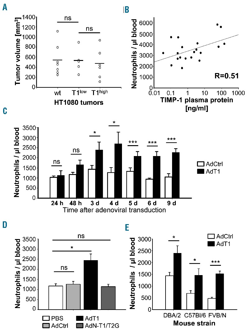
Figure 1.

TIMP-1 plasma levels correlate with increased neutrophil counts in peripheral blood. (A) Volume of HT1080 tumors expressing different TIMP-1 levels. 2*106 tumor cells were s.c. injected into the neck of CD1nu/nu mice and allowed to grow over ten days. (B) Correlation of TIMP-1 plasma levels with neutrophil blood counts in mice from (A). (C) Neutrophil blood counts in DBA/2 mice transduced to express high TIMP-1 levels. TIMP-1 coding (AdT1) or control (AdCtrl) adenovirus was i.v. injected into mice and peripheral blood neutrophils were quantified by flow cytometry at the indicated time points. (D) Neutrophil blood counts in DBA/2 mice three days after injection of adenovirus or PBS. (E) Neutrophil blood counts in different mouse strains three days after adenovirus injection. Columns: mean; Bars: SEM; Student’s t-test. ns: not significant; *P<0.05; ***P<0.001, n=5.
