Figure 2.
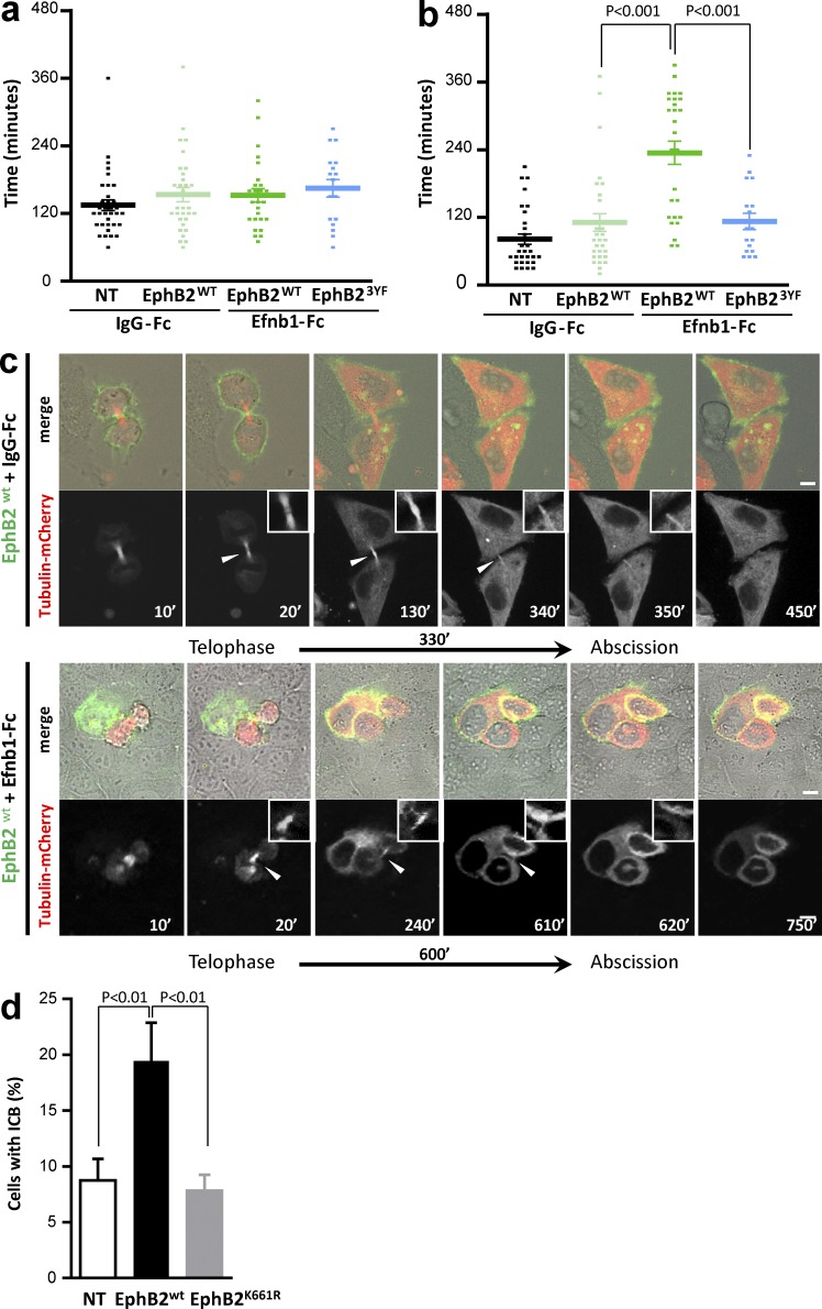
EphB2-induced abscission defect is kinase dependent. (a and b) HeLa cells were left untransfected (NT) or were transfected with EphB2WT-GFP (EphB2WT) or EphB23YF-GFP (EphB23YF) for 48 h and synchronized, and cell division was recorded with an Incucyte microscope, in conditions of mock treatment (IgG-Fc) or Efnb1-Fc treatment. (a) Duration of the progression from anaphase to telophase in IgG-Fc– or Efnb1-Fc–treated cells. (b) Duration of the progression from telophase to disappearance of the ICB in IgG-Fc– or Efnb1-Fc–treated cells. (c) Still images from confocal recordings of dividing cells in EphB2WT-GFP cells expressing tubulin-mCherry mock treated or treated with Efnb1-Fc (as indicated). The top rows show fluorescence and phase contrast overlay, and the bottom rows show the tubulin-mCherry fluorescence to highlight the ICB (arrowhead). Insets are higher-magnification images of the ICB. (d) HeLa cells either NT or transfected with EphB2WT or EphB2K661R were treated with Efnb1-Fc. The proportion of cells connected by an ICB after 24 h of treatment was quantified in each condition. Error bars correspond to SEM. Statistical p-value is indicated when significant. Bars, 10 µm.

