Figure 6.
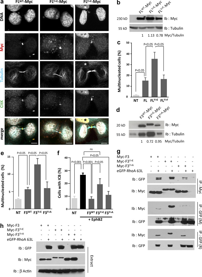
Tyrosine phosphorylation of CitK is detrimental to abscission. (a) HeLa cells were transfected with Myc-tagged CitK FL, phosphomimetic CitK (FLY>E), or unphosphorylatable CitK (FLY>F), fixed, and immunostained to detect Myc (red), tubulin (cyan), or CitK (green). Nuclei were stained with DAPI (gray). Nuclear localization of CitK (asterisks) corresponds to interphase cells. Bars, 10 µm. (b and c) HeLa cells were untransfected (NT) or transfected for 48 h with Myc-tagged CitK FL, phosphomimetic CitK (FLY>E), or unphosphorylatable CitK (FLY>F). (b) Protein extracts were immunoblotted with anti-myc and -tubulin antibodies. Ratio of signal intensity is provided below. (c) The proportion of multinucleated cells was quantified in each condition. (d and e) HeLa cells were untransfected (NT) or transfected for 48 h with Myc-tagged CitK-F3 (F3WT), phosphomimetic CitK-F3 (F3Y>E), or unphosphorylatable CitK-F3 (F3Y>A) fragments. (d) Protein extracts were immunoblotted with anti-myc and -tubulin antibodies. Ratio of signal intensities is indicated below. (e) The proportion of multinucleated cells was quantified in each condition. (f) HeLa cells were untransfected (NT) or cotransfected for 48 h with EphB2 alone (black bar) or Myc-tagged CitK-F3WT, phospho-mimetic CitK-F3Y>E, or unphosphorylatable CitK-F3Y>A fragments. Cells were stimulated with Efnb1-Fc for 24 h, and the proportion of cells connected by an ICB was quantified in each condition. (g and h) HEK-293 cells were transfected with Myc-tagged CitK-F3WT (F3), phospho-mimetic CitK-F3Y>E (F3Y>E), or unphosphorylatable CitK-F3Y>A (F3Y>A) and an activated form of RhoA tagged with GFP (eGFP-RhoA63L) as indicated. (g) CitK fragments were immunoprecipitated (top, Ip Myc) with an anti-Myc antibody and immunoblotted with GFP and Myc antibodies. RhoA was immunoprecipitated (middle and bottom, Ip GFP) with anti-GFP antibodies of different species and immunoblotted with GFP and Myc antibodies. (h) Protein lysates (Extract) were probed with antibodies to detect GFP and Myc. Actin was used as loading control. Western blots are representative of at least two independent experiments. Error bars correspond to SEM. Statistical p-value is indicated when significant. ns, nonsignificant.

