Figure 1.
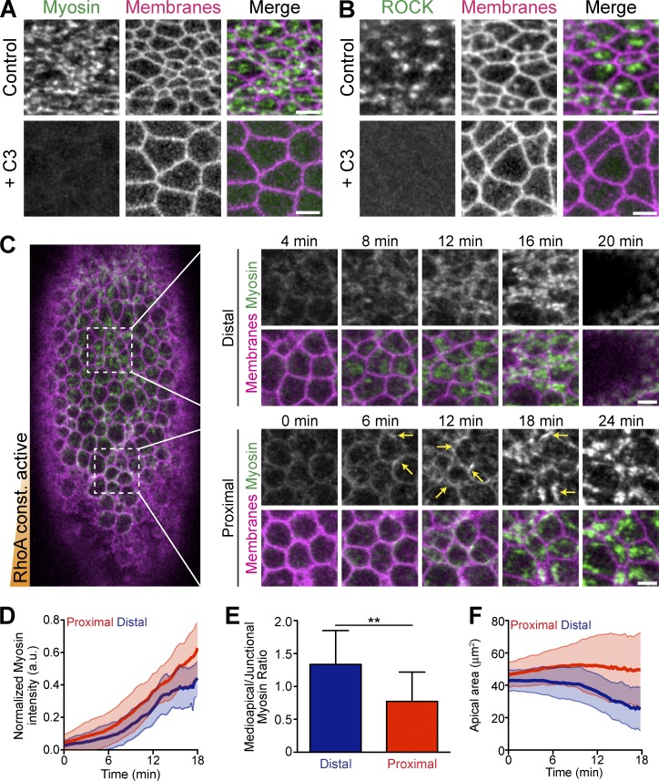
Constitutively active RhoA is insufficient for apical constriction. (A) Apical myosin depends on RhoA activity. Images from live embryos expressing myosin::GFP (sqh::GFP) and membrane::RFP (Gap43::mCherry) and injected with buffer (PBS, Control) or the C3 exoenzyme immediately before gastrulation. (B) Apical ROCK localization depends on RhoA activity. Images from live embryos expressing GFP::ROCK and Membrane::RFP injected with solvent or C3 immediately before gastrulation. (C) Constitutively active RhoA (CA-RhoA) disrupts proper myosin organization in ventral furrow cells. Image of a live embryo expressing Myosin::GFP and Membrane::RFP that has been injected at one pole (bottom) with mRNA encoding CA-RhoA (G14V). Time-lapse images (right) show myosin accumulating abnormally at junctions (arrows), specifically in cells proximal to injection. (D) Quantification of total apical myosin intensity in embryo in C injected with CA-RhoA mRNA (n = 47 cells proximal, 44 cells distal from two embryos; error bars represent ±SD). Note that myosin intensity is slightly higher close to the injection site (proximal). a.u., arbitrary units. (E) Quantification of myosin intensity in the middle of the apical surface (medioapical) relative to the junctions. Cells proximal to CA-RhoA injection have a lower medioapical-to-junctional ratio (n = 12 cells proximal, 12 cells distal; **, P < 0.01, unpaired t test; error bars represent ±SD). (F) Apical constriction is impaired in cells proximal to CA-RhoA injection (n = 47 cells proximal, 44 cells distal; error bars represent ±SD). Bars, 5 µm.

