Figure 2.
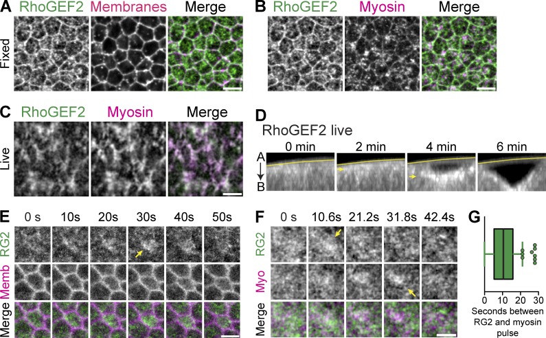
Medioapical RhoGEF2 pulses precede ROCK-myosin. (A–C) RhoGEF2 localizes across the medioapical surface. (A and B) Images from fixed Myosin::GFP (sqh::GFP) embryos, stained with a RhoGEF2 antibody. Membranes are subapical F-actin (phalloidin). (C) Images from live embryos expressing GFP::RhoGEF2 and myosin::RFP. Note the strong colocalization of RhoGEF2 with myosin. (D) Images of cross section views of ventral furrow formation in live embryos showing increasing apical accumulation of GFP::RhoGEF2 (arrows). Line marks the vitelline membrane. (E) Time-lapse images represent apical GFP::RhoGEF2 and subapical membrane::RFP. Pulses of RhoGEF2 occur across the entire medioapical region (arrow). (F) Time-lapse images of embryo expressing GFP::RhoGEF2 and myosin::RFP show pulsed accumulation (arrows). (G) Quantifications of time delay, in seconds, between RhoGEF2 and myosin pulses (mean = 10.4 ± 7 s, n = 187 pulses, 5 embryos; error bars represent SD). Bars: (A–C) 5 µm; (E and F) 2.5 µm.

