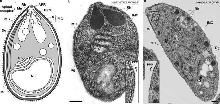
Figure 1.
Motile apicomplexan zoite cells designed for invasion and motility. (a) Schematic of an apicomplexan zoite cell (here a merozoite) showing key cellular structures and apical complex, characteristic of motile zoites. (b and c) Electron micrographs of a P. knowlesi merozoite (b) and a Toxoplasma tachyzoite (c). Apicomplexan zoites are generally polarized and elongated, with either a crescent or oval shape. Each has a distinctive apical complex, which consists of secretory organelles called micronemes, rhoptries, and dense granules. Micronemes (oval or pear-shaped organelles) secrete their contents at the anterior tip of motile zoites during motility/invasion. Rhoptries (club-shaped organelles) fuse and release their contents concomitantly with host-cell invasion (Carruthers and Tomley, 2008; Counihan et al., 2013; Hanssen et al., 2013). Dense granules (a mixed grouping of secretory vesicles) are released via fusion with the plasma membrane before or after invasion (also called exonemes; Yeoh et al., 2007). Insets highlight the triple-layered appearance of the parasite pellicle at higher magnification (double-membraned IMC, lying under the PPM). The myosin motor is thought to lie between the outer (o) IMC membrane and the PPM. APR, apical polar (tubulin-rich) rings; Dg, dense granules; Go, Golgi apparatus; i, inner membrane of the IMC; Mn, micronemes; Mt, subpellicular microtubules; Nu, nucleus; Rh, rhoptries. Bars, 200 nm. Micrograph images courtesy of L.H. Bannister (Kings College London, London, England, UK) and D. Ferguson (University of Oxford, Oxford, England, UK).

