Fig. 4.
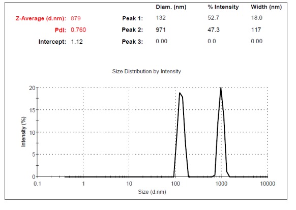
Fig. 4a .

Dynamic Light Scattering (DLS) results for the size distribution of MVs obtained at the centrifugation rate of 10,000× g. From the relative intensities of the size distribution peaks, appears that there is likely some apoptotic debris within the sample.
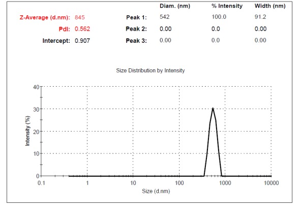
Fig. 4b .

Dynamic Light Scattering (DLS) results for the size distribution of MVs obtained at the centrifugation rate of 20,000× g.
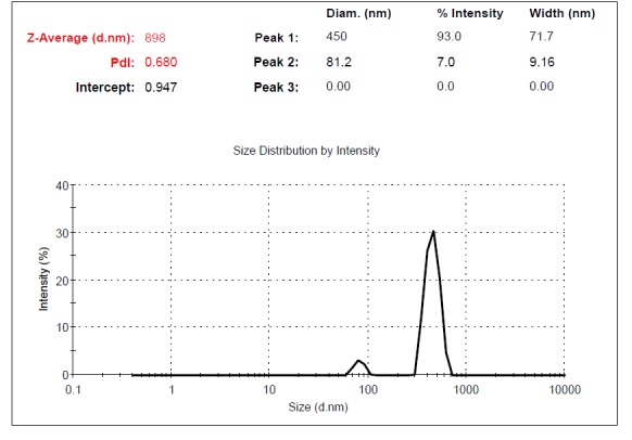
Fig. 4c .

Dynamic Light Scattering (DLS) results for the size distribution of MVs obtained at the centrifugation rate of 40,000× g. The contamination possibility with exosomes at 40,000× g is less than at 60,000× g.
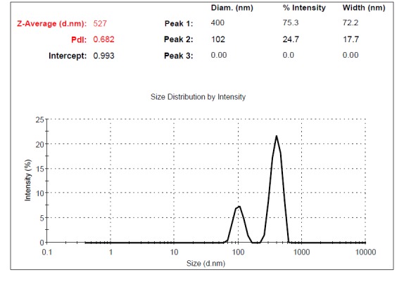
Fig. 4d .

Dynamic Light Scattering (DLS) results for the size distribution of MVs obtained at the centrifugation rate of 60,000× g.
