Fig. 3.
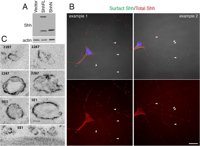
Shh immunolabeled EVs are found in primary cultures of hippocampal neurons. (A) Validation of the specificity of Shh 2287 antibody. ShhFL, a full-length Shh construct; ShhN, an N-terminal Shh fragment construct; Vector, control vector. (B) Representative confocal images of cultured hippocampal neurons labeled with two different Shh antibodies. Cell surface Shh was detected by labeling live neurons with one Shh antibody (2287, green); total Shh was detected by labeling fixed and permeabilized neurons with another Shh antibody (5E1, red). Note that in addition to punctate labeling on neurons, many Shh-labeled puncta are seen outside of and away from neurons (white arrowheads). Images are representative of data observed in at least four separate cultures. Scale bar: 20 µm. The controls for this labeling experiment are shown in Fig. S2. (C) Representative electron micrographs of Shh-labeled EVs found in primary cultures of hippocampal neurons. Neurons were fixed and labeled with either Shh 5E1 antibody or Shh 2287 antibody, followed by a biotinylated secondary antibody and avidin-biotin-peroxidase reagent before processing for electron microscopy. Dark surface labeling is the peroxidase-DAB reaction product. Notice that Shh-labeled EVs have a wide range of sizes. Scale bar: 100 nm.

