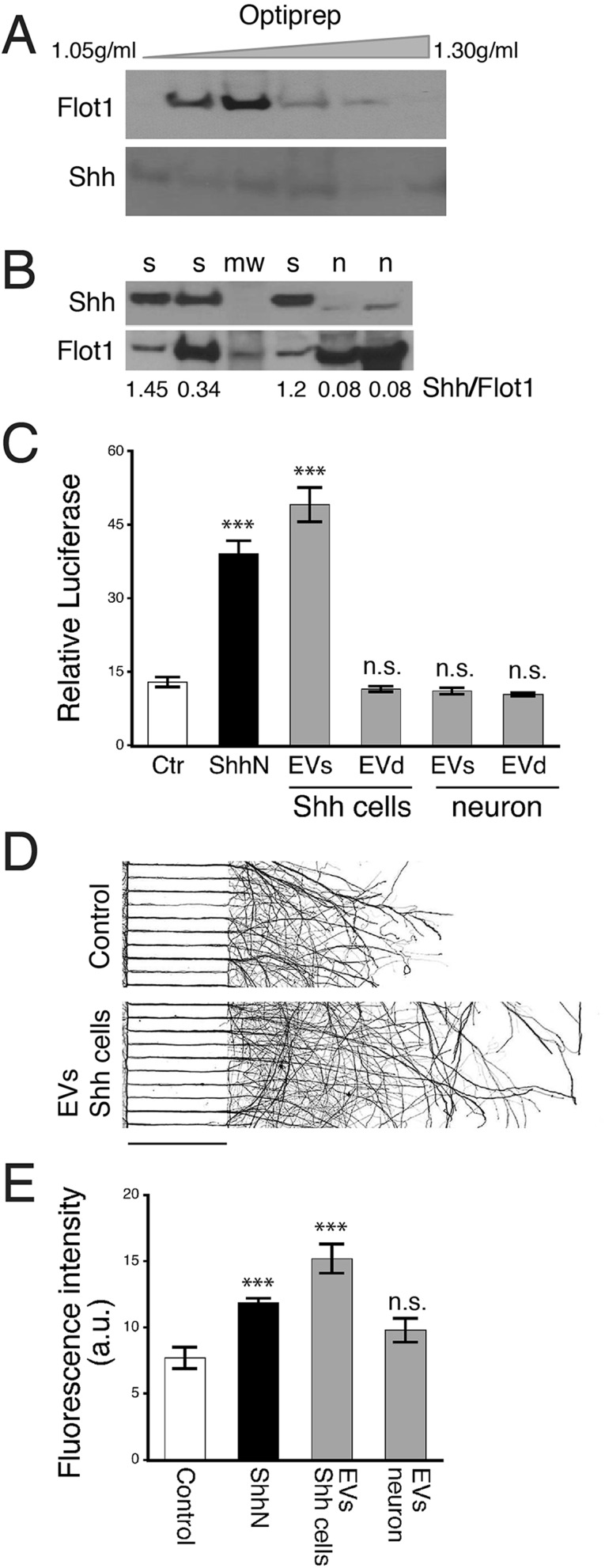
Fig. 4.
Biochemical and functional measures of EVs isolated from cultured cells. (A) EVs were isolated from the culture medium of a Shh-overexpressing cell line, ShhN-HEK293 (Shh cells). Immunoblots of density-fractionated samples show that Shh (detected by Shh 2287 antibody) is present, but is not concentrated in the Flot1-enriched exosome fraction. (B) EVs isolated from culture medium of hippocampal neurons were subjected to immunoblot analysis. Quantification of Shh by normalizing to Flot1 show significantly smaller amounts of Shh in neuron-derived EVs compared to EVs derived from Shh-overexpressing cells. s, Shh-overexpressing cells; n, neurons; mw, molecular weight markers. (C) Shh bioactivity was measured using Shh-Light2 assay. EVs from the Shh-overexpressing cell line (Shh cells) displayed high Shh activity, whereas EV-depleted samples (EVd) had no Shh activity. EVs from neurons were also assayed. No Shh bioactivity was detected in either EVs or EVd samples. Control (Ctr), culture medium from HEK293 cells; ShhN, culture medium from ShhN-HEK293 cells (5%). The assay was repeated three times. Data represented as mean±s.e.m.; ***P<0.001; n.s., not significant. (D) Representative images of neurons grown in a compartmentalized culture system for 6 days. EVs isolated from ShhN overexpressing cells were added to the soma compartment, and neurons were grown for one additional day. Neurons were immunolabeled with a neuronal marker, Tuj1. Images were inverted for better visualization of axons. Scale bar: 450 µm. (E) Quantitative analysis of Tuj1 fluorescence intensity in axons. The experiment was repeated four times. Data represented as mean±s.e.m.; ***P<0.001; n.s., not significant.

