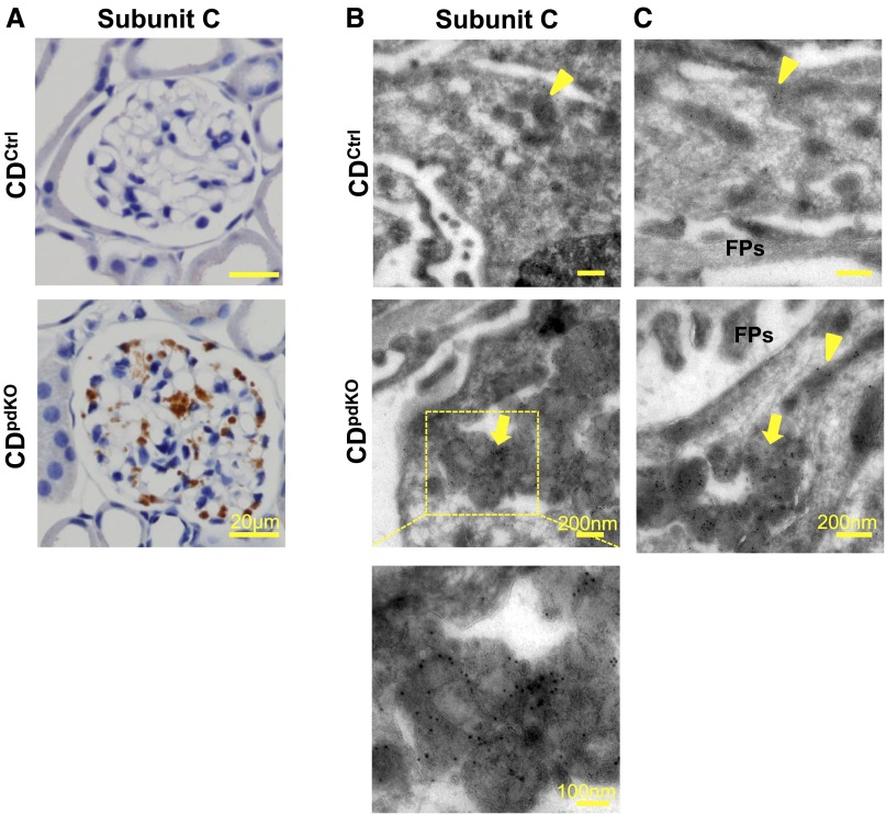
Figure 7.
Subunit c of mitochondrial ATP synthase accumulate in lysosomal structures of podocytes as in CD-deficient neurons. (A) Immunohistochemical staining showed subunit c of mitochondrial ATP synthase accumulated in 10-month-old CDpdKO mouse podocytes. (B and C) Immunoelectron microscopy indicated that, in podocytes from CDpdKO mice, the labeling for subunit c was associated with membrane-bound structures containing GRODs. Immunocytochemical staining of subunit c of mitochondrial ATP synthase in podocytes from CDpdKO mice and their CDCtrl littermates at 10 months using the cryothin section immunogold method is shown. (B, top panel and C, upper panel) Subunit c gold particles label only the mitochondrial inner membrane in the CDCtrl littermates (arrowheads), whereas (B, middle panel and C, lower panel) they are associated with both the inner membrane of intact mitochondria (arrowhead) and the membrane-bound compartments with dense materials (GRODs; arrows) in the CDpdKO mice.

