Figure 8.
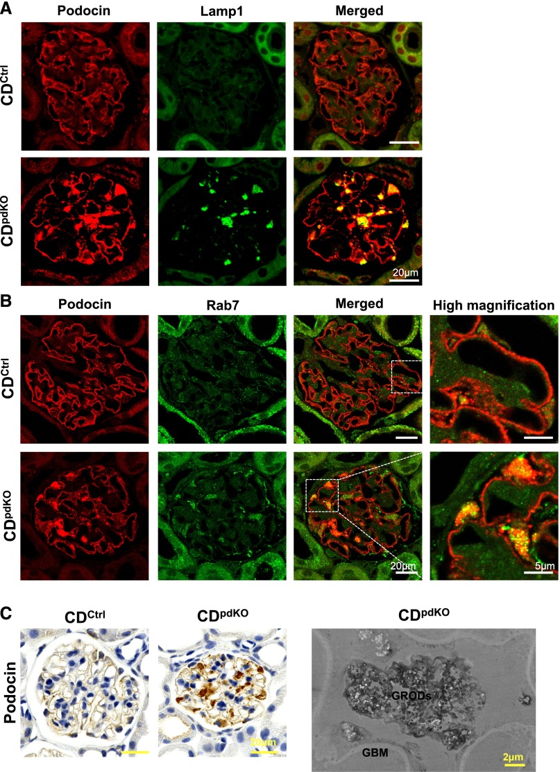
The accumulation of podocin in 10-month-old CDpdKO mouse podocyte cell bodies is particularly colocalized with Lamp1 and Rab7. (A and B) Immunofluorescence staining showed the localization of podocin in the GBM in CDCtrl littermates. By contrast, podocin was localized in both the GBM and podocyte cell bodies in CDpdKO mice. The late endosomal marker Rab7 was increased in CDpdKO mouse podocytes compared with CDCtrl littermate podocytes. Podocin localized in podocyte cell bodies was particularly colocalized with (A) Lamp1 and (B) Rab7 in CDpdKO mice. (C) For immunohistochemical staining, we detected a strong accumulation of podocin in CDpdKO mouse podocyte cell bodies. Immunoelectron microscopy using paraffin–embedded tissue sections indicated that the granular structures (GRODs) that accumulate in the podocyte cell bodies of CDpdKO mouse glomeruli are autophagosomes/autolysosomes containing degraded podocin.

