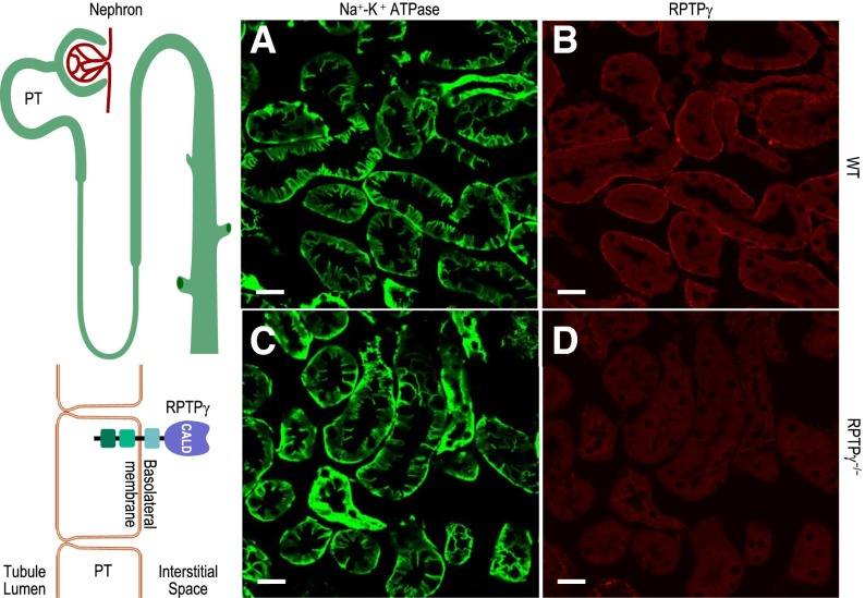
Figure 1.
PTs express RPTPγ on the portion of the basalateral membrane that faces blood vessels. (A–D) Immunofluorescent staining of cortical sections of kidneys from WT or RPTPγ−/− mice, showing predominantly PTs. The sodium-potassium adenosine triphosphatase (Na+-K+ ATPase) antibody (green, BL membrane marker) stains all regions of the BL membrane in PTs from WT and RPTPγ−/− mice. Our RPTPγ antibody (red) establishes that RPTPγ protein was present only in the subset of the PT BL membrane facing the blood vessels in WT PTs. Images are representative of data obtained from three mice per group. Scale bar =20 μm.

