Figure 2.
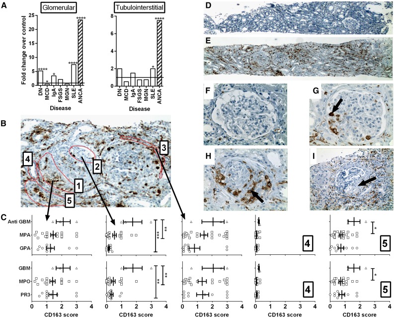
CD163 is highly expressed in the kidneys of patients with vasculitis. (A) RNA was extracted from microdissected glomerular and tubulointerstitial compartments from patients with diabetic nephropathy (DN), minimal change disease (MCD), IgA nephropathy (IgA), FSGS, MGN, lupus nephritis (SLE), and ANCA vasculitis. The degree of expression of the CD163 gene compared with microdissected healthy control kidney was determined by Affymetrix microarrays. Bars represent fold changes compared with the respective controls. (****q<0.01%; q<5%) (b) Paraffin-embedded human kidney sections from patients with vasculitis were stained for CD163 protein by immunohistochemistry and scored blind according to the location of cells with each of five regions: (1) within regions of fibrinoid necrosis or crescent formation, (2) within regions of apparently normal glomeruli, (3) in the periglomerular region, (4) within tubules, and (5) in the interstitial compartment. (C) CD163 scores in each of the respective five regions stratified by clinical diagnosis (upper graphs) and antibody specificity (lower graphs), (P<0.05; **P<0.01; ***P<0.001). (D–I) Images depict representative low power (×40 magnification) views of healthy control (D) and vasculitic (E) kidney, alongside high power (×400) views of healthy control kidney (F), a glomerulus with mild vasculitic injury (G, arrow), a severely affected glomerulus with established crescent formation (H, arrow), and a glomerulus with a fibrous crescent from previous vasculitic injury (I, arrow, ×200). MPA, microscopic polyangiitis; GPA, granulomatosis with polyangiitis.

