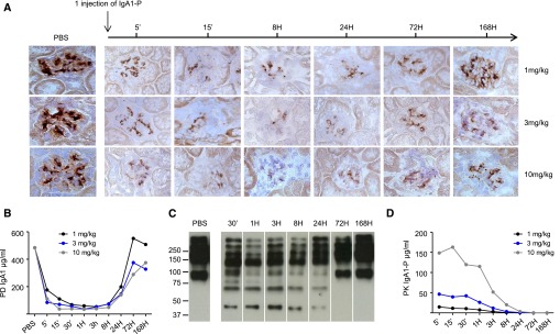
Figure 1.
Decrease in serum IgA1 levels and mesangial IgA1 deposits after IgA1‑P treatment. (A) IgA1 deposit detection in the mesangium (brown) after IgA1‑P IV injection (1, 3, and 10 mg/kg) in α1KI-CD89Tg mice (n=3 mice per group) using immunohistochemistry directed against human IgA on frozen kidney sections. Representative sections. (B) Concentrations of serum IgA1 after IgA1‑P injection for each time point of euthanized mice as described in (A). (C) Detection of IgA1 fragments by western blot in serum from mice after 3 mg/kg injection. Representative samples of selected time points. (D) Pharmacokinetics (PK) of IgA1‑P in serum of α1KI-CD89Tg mice after IV injection for each time point of euthanized mice as described in (A). PD, pharmacodynamics.

