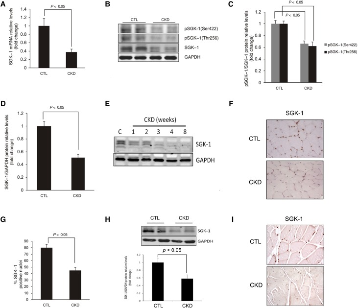
Figure 1.
CKD suppresses SGK-1 expression and activation. TA muscles of CKD and pair–fed control mice were analyzed by (A) RT-PCR and (B) Western blot. (C and D) The intensity analysis of SGK-1 protein and its phosphorylation are summarized. (E) TA muscle were collected from mice at different time points after the removal of the second kidney, and the expression of SGK-1 protein was determined. (F) Immunohistologic staining of SGK-1 in TA muscle (brown nuclei are SGK-1 positive). (G) The percentages of SGK-1–positive nuclei in TA muscles of control or CKD mice are shown. (H and I) SGK-1 expression in patients with CKD was detected by (H) Western blot and (I) immunostaining; the density analysis was performed and is shown in H, lower panel. For A, C, D, and G, five mice in each group were examined; for E, three mice in each group were examined (P<0.05). CTL, control; GAPDH, glyceraldehyde-3-phosphate dehydrogenase.

