Figure 2.
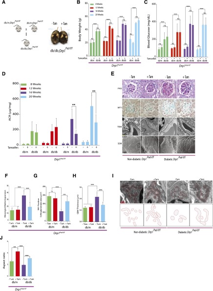
Conditional deletion of Drp1 in podocytes protects against progression of DN. (A, left panel) Mating strategy to generate Drp1 conditional knockout in mouse podocytes of diabetic mice. (A, right panel) Representative image of tamoxifen–induced and noninduced diabetic db/db;Drp1Pod-f/f mice. (B) Body weight, (C)blood glucose levels, and (D) albumin-to-creatinine ratios (ACRs; micrograms per milligram) of tamoxifen-induced and noninduced controls from nondiabetic (db/m;Drp1Pod-f/f) and diabetic (db/db;Drp1Pod-f/f)mice at 4-week intervals beginning at 8 weeks of age (n=10 mice per group).(E) Representative micrographs of PAS–stained kidney sections (row 1), WT1 (row 2), transmission electron micrographs (row 3), and scanning electron micrographs (row 4) from the groups described in D. Scale bars, 10μm for rows 1 and 2; 500nm for row 3; 2 μm in row 4. (F) Quantification of mesangial matrix expansion (n=10 per group) and (G) WT1-positive glomeruli from 25 randomly selected glomeruli per animal in each indicated group (n=5 mice per group). (H) Quantification of GBM thickness (n=5–7 mice per group). (I, upper panel) Representative TEM micrographs of podocyte mitochondria from groups described in D. (I, lower panel) Tracing of mitochondria from aforementioned TEM micrographs. (J) Average aspect ratios in each group. Results are presented as means±SEMs. **P<0.01; ***P<0.01; ****P<0.001.

