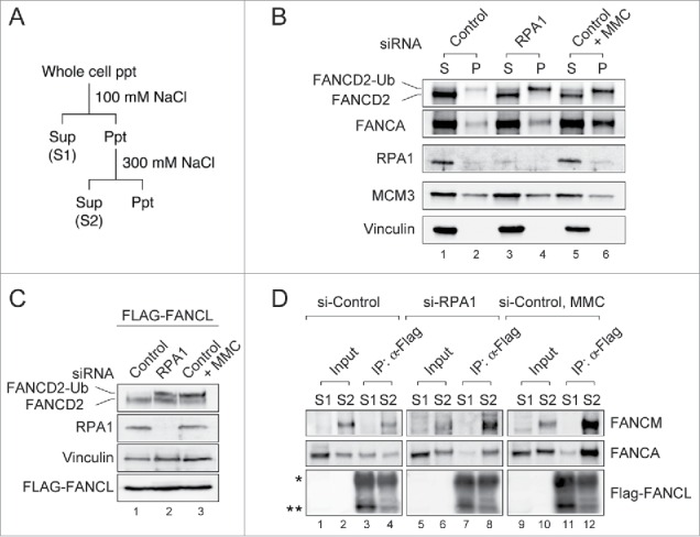
Figure 3.

RPA1 depletion increases the chromatin association of FA core complex. (A) Experimental protocol used to prepare cell extracts described in panel B-D. (B) U2OS cells were transfected with either control or RPA1-specific siRNA. 72 hr after transfection, cells were fractionated into S (Sup) & P (Ppt) and immunoblotted for the indicated proteins. MCM3 and Vinculin were used as loading controls. (C-D) HeLa cells expressing FLAG-tagged FANCL were transfected with either control siRNA or RPA1-specific siRNA and cultured for 72hr. (C) Whole-cell lysates were prepared and analyzed with indicated antibodies. (D) Cells were fractionated into S1 and S2. Each fraction was immunoprecipitated with FLAG-M2 agarose beads, and the precipitates were immunoblotted with the indicated antibodies. The asterisk indicates the IgG heavy chain and the double asterisk shows the position of FLAG-tagged FANCL. Entire images come from the identical gel.
