Figure 5.
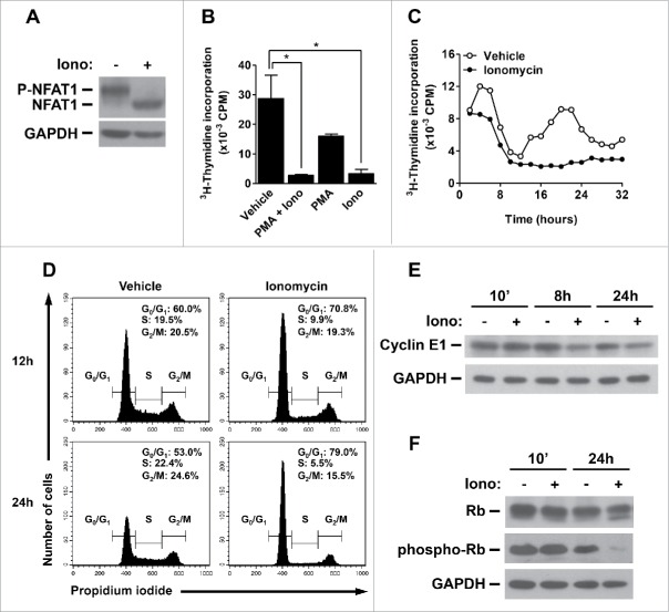
NFAT1 induces cell cycle arrest and inhibits Cyclin E expression in B cell lymphoma. (A) Total protein lysates were obtained from Raji cells stimulated or not with Ionomycin (5 μM) in the presence of 2 mM of CaCl2 for 10 minutes. NFAT1 and GAPDH protein levels were detected by protein gel blot. (B) Raji cells were stimulated or not with PMA (10 nM) and/or Ionomycin (5 μM) for 24 hours. (C) Raji cells were stimulated or not with Ionomycin (5 μM) for 32 hours and analyzed every 2 hours. (B and C) After stimulation, cells were pulsed with 3H-thymidine (5 μCi/mL) for 2 hours. Cells were then harvested and 3H-thymidine incorporation was analyzed by β-spectrometer. CPM refers to counts per minute. (D) Raji cells were stimulated or not with Ionomycin (5 μM) for the indicated time points. Cells were then labeled with propidium iodide, and DNA content was analyzed by flow cytometry for cell cycle phases. (E and F) Total protein lysates were obtained from Raji cells stimulated or not with Ionomycin (5 μM) for the indicated time points. Cyclin E1, Rb, phospho-Rb (Thr821 and Thr826), and GAPDH protein levels were detected by western blot. All results are representative of at least 3 independent experiments. Results are expressed as mean and error bars represent SD. Asterisks indicate significance levels compared to controls (p < 0.05).

