Figure 1.
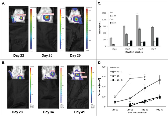
Mice were imaged via bioluminescent imaging throughout disease progression to monitor development and spread of metastases. Mice in the ad lib (A) fed group developed visible metastases significantly faster than those in the CR fed groups (B). Imaging intensity is designed such that it is proportional to the proliferative capacity of the cells in the luminescent area. On average, metastases in the AL fed groups demonstrated increased intensity and were larger than those in the CR fed groups. The last imaging time point for AL fed mice was day 29 compared with day 41 for the CR fed group. Total volume of metastatic disease burden within the lungs was calculated for each treatment group at each respective time point (C). AL fed mice had larger total volume across time points when compared with other treatment groups, specifically at later time points as metastatic disease volume approached 1400 mm3. Average size of lung nodules on imaging was calculated for each imaging time point as well (E). AL fed mice had significantly larger lung nodules on average across imaging time points. Total volume of metastases and average nodule size were based on a representative 3 mice from each cohort, where error is defined as SEM with n = 3.

