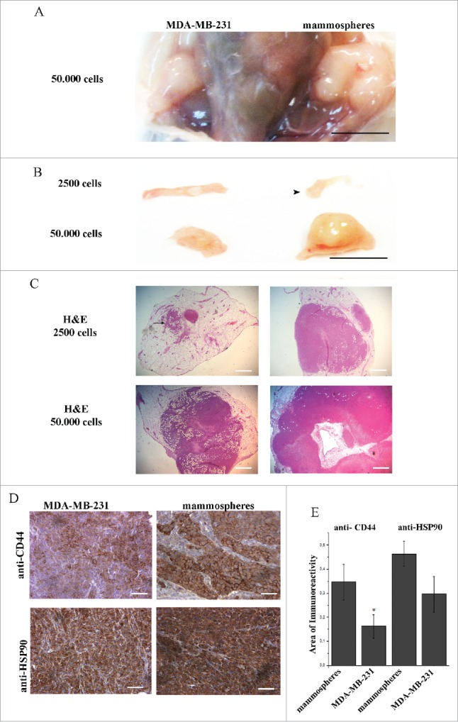
Figure 4.

Orthotopic inoculation of MDA-MB-231 cells and mammospheres, revealed enrichment of CD44 positive cells in mammosphere-derived tumors. Eight weeks post inoculation, left and right panels show MDA-MB-231- and mammosphere-derived tumors, respectively. (A) The anatomical site demonstrating tumors formed in both mammary fat pads after inoculation of 50,000 cells/site. (B) Macroscopic pictures of orthotopic xenografts. Arrowhead shows a palpable mammosphere-derived tumor after injection of 2,500 cells. (C) Corresponding hematoxylin/eosin (H&E) stained sections of the xenografts shown in (B). Administration of 2,500 MDA-MB231 cells results in a very small niche of cancer cells (arrow). (D) Immunostaining of tumors derived from mice injected with 50,000 cells/site, using anti-CD44 and anti-HSP90 antibodies. (E) Quantification of CD44 expression showed a significant enhancement (*p < 0.05) in the mammosphere-derived tumors as compared to the MDA-MB-231-derived ones, whereas increased HSP90 expression in the mammosphere-derived tumors as compared to the MDA-MB-231 derived ones was not statistically significant. *p < 0.05. In (A) and (B) bars correspond to 10 mm, in (C) bars correspond to 1100 μm and in (D) bar corresponds to 100 μm.
