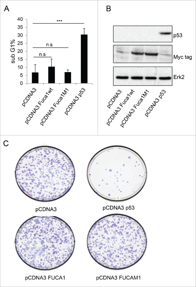
Figure 4.

FUCA1 overexpression is not sufficient to induce cell death. Saos 2 cells were transiantly co-transfected with either an empty pcDNA3 vector, a wild-type-Fuca1wt (pCDNA3 FUCA1wt), an enzymatically inactive mutant of FUCA1 (pCDNA3 FUCA1M1), or a wild-type-p53-expressing construct together with pCMV-CD20. After 72 h, the transfected cells were identified by staining for CD20 and analyzed for sub-G1 DNA content by flow cytometry (n = 4 independent experiement, one way Anova ***p < 0.0001) (A). (B) 24 hours post transfection p53 and Fuca1 expression were assessed by western blotting using respectively an anti-p53 antibody and an anti-Myc-tag antibody. Immunoblot against actin was used as a loading control. (C) Saos-2 cells were transfected with either an empty pcDNA3 vector, a wild-type-FUCA1wt (pCDNA3 FUCA1wt), an enzymatically inactive mutant of FUCA1 (pCDNA3 FUCA1M1), or a wild-type-p53-expressing construct. Following selection, cells were assessed for clonogenic survival using Giemsa staining (C).
