Figure 3.
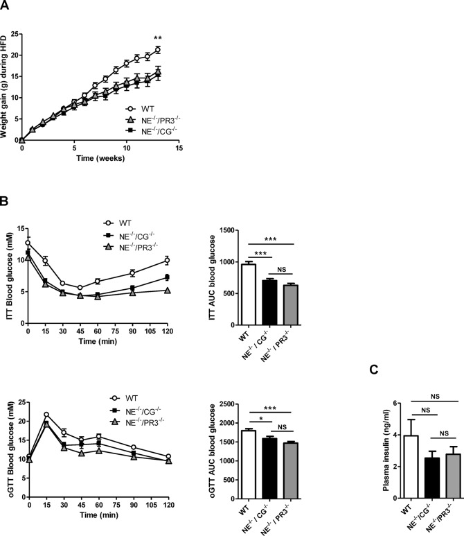
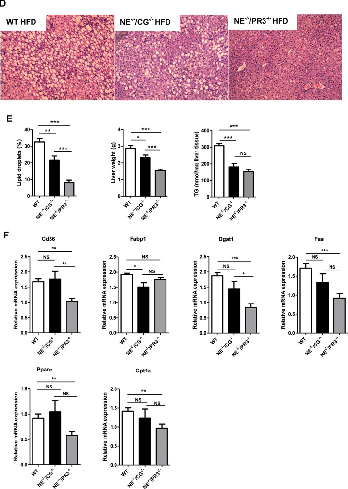
Improved NAFLD status and metabolic profiles in NE–/–/CG–/– and NE–/–/PR3–/– mice after a high-fat diet. (A) Bodyweight development of wild-type (n = 10), NE–/–/CG–/– (n = 15) and NE–/–/PR3–/– (n = 15) mice during diet intervention. (B) Insulin tolerance test and ITT AUC values, oral glucose tolerance test and oGTT AUC values of wild-type, NE–/–/CG–/– and NE–/–/PR3–/–. (C) Plasma insulin levels. (D) HE staining of liver sections. (E) Percentage of lipid droplets in HE-stained liver sections, liver weights and liver triglycerides levels. (F) Relative mRNA expression of Cd36, Fabp1, Dgat1, Fas, Ppar α and Cpt1a in liver. HFD, high-fat diet; WT, wild-type; ITT, insulin tolerance test; oGTT, oral glucose tolerance test; TG, triglycerides; *significant difference p ≤ 0.05; **significant difference p ≤ 0.01; ***significant difference p ≤ 0.001.


