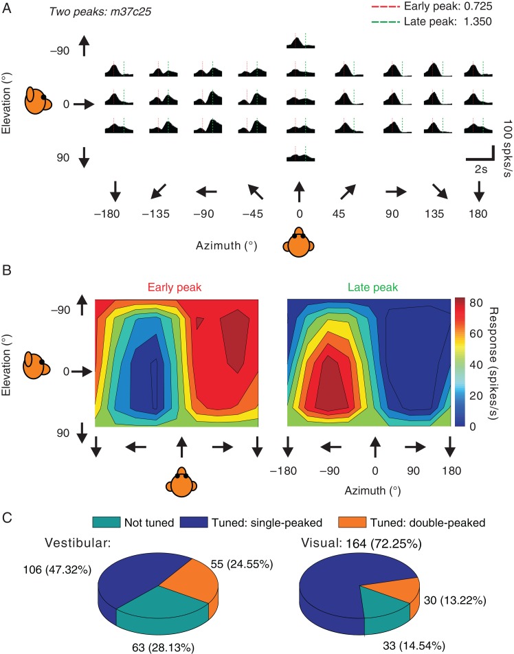
Figure 2.
Double-peaked example neuron and summary of response dynamics. (A) Example of an FEFsem neuron with double-peaked heading tuning in the vestibular condition. PSTHs are shown for 26 directions of vestibular translation. The red and green dashed vertical lines indicate the 2 peak times. (B) 3D heading tuning of the same example neuron at each of the 2 (early and late) peak times. Spikes were counted during a 400 ms window around each peak time. (C) Pie charts for the vestibular (left) and visual (right) conditions, showing the proportions of neurons in FEFsem with significant single-peaked tuning and double-peaked tuning, as well as neurons that were not tuned.

