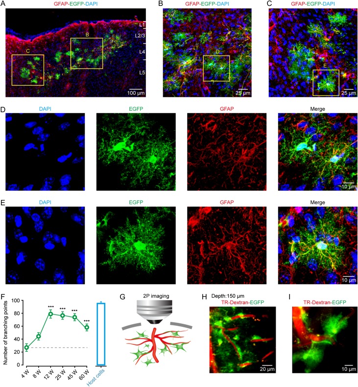
Figure 2.
Engrafted astrocytes are morphologically integrated into the adult mouse cortex and survive for at least 60 weeks. (A) Representative image of engrafted astrocytes in a cortical region at 12 weeks after transplantation. Engrafted astrocytes, positive for EGFP/GFAP, were distributed in different cortical layers. DAPI was used to stain nuclei throughout the study.(B–E) Higher magnification of regions from panel A showing the complex fine structures of engrafted astrocytes (EGFP+). (F) Summary of the number of processes of engrafted astrocytes at different weeks (W) after transplantation (green points), and of their host astrocytes (host cells, blue bar graph). The host cells are those GFP-negative but GFAP-positive cells from the same preparations. The number of processes is quantified by the defined branching points. n = 20–21 cells from 4 to 9 mice for each group; ***P < 0.001 versus 4 W, Student's t test. (G) Cartoon depicting the relative position between the processes of engrafted astrocytes and blood vessels under in vivo two-photon imaging setup. (H, I) Representative images of engrafted astrocytes (EGFP+) wrapping and making contacts with blood vessels 12 weeks after transplantation. The vasculature was visualized using Texas Red-dextran (TR-Dextran). The depth from pial surface of the imaging is 150 µm.

