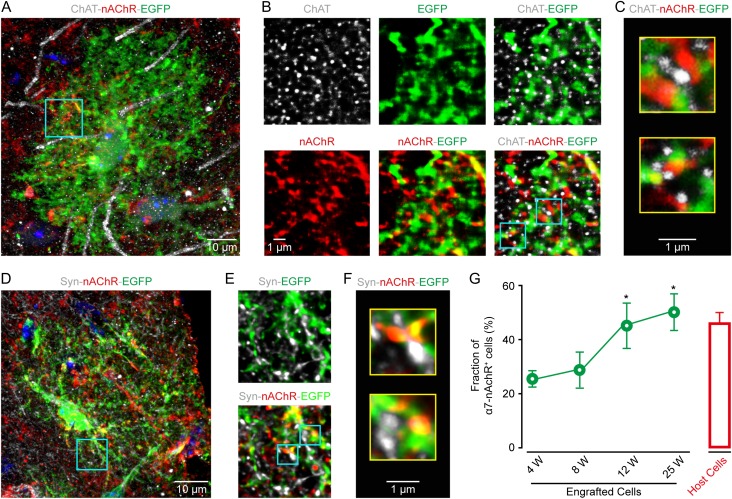
Figure 4.
Engrafted astrocytes express nicotinic acetylcholine receptors. (A) Immunostaining image of EGFP (green) for engrafted astrocytes, ChAT (gray) for cholinergic structures, and nAChR α7 (red) in the somatosensory cortex at 12 weeks after transplantation. (B) High magnification of the region outlined in panel A. (C) Higher magnification of two regions outlined in panel B, showing engrafted astrocytic processes (EGFP+), often expressing nAchRs and next to presynaptic cholinergic puncta (ChAT+). (D) Confocal image of synaptophysin (syn, gray), nAchR (red), and EGFP (green) in a transplanted cortical region at post-transplantation week 12. (E) High magnification of the expression of Syn, EGFP and α7 of the region outlined in panel D. (F) Higher magnification of two regions outlined in panel E. nAchR labeling decorates engrafted astrocytic processes (EGFP+), often facing syn-labeled (Syn+) presynaptic elements. (G) Fraction of nAchR+ engrafted astrocytes (green) at different weeks after transplantation (n = 4–7 sections, 41–150 cells from 4 mice for each group), and of their host astrocytes (red, n = 4 sections, 72 cells). Mann–Whitney U test, *P < 0.05 versus 8 W.

