Figure 7.
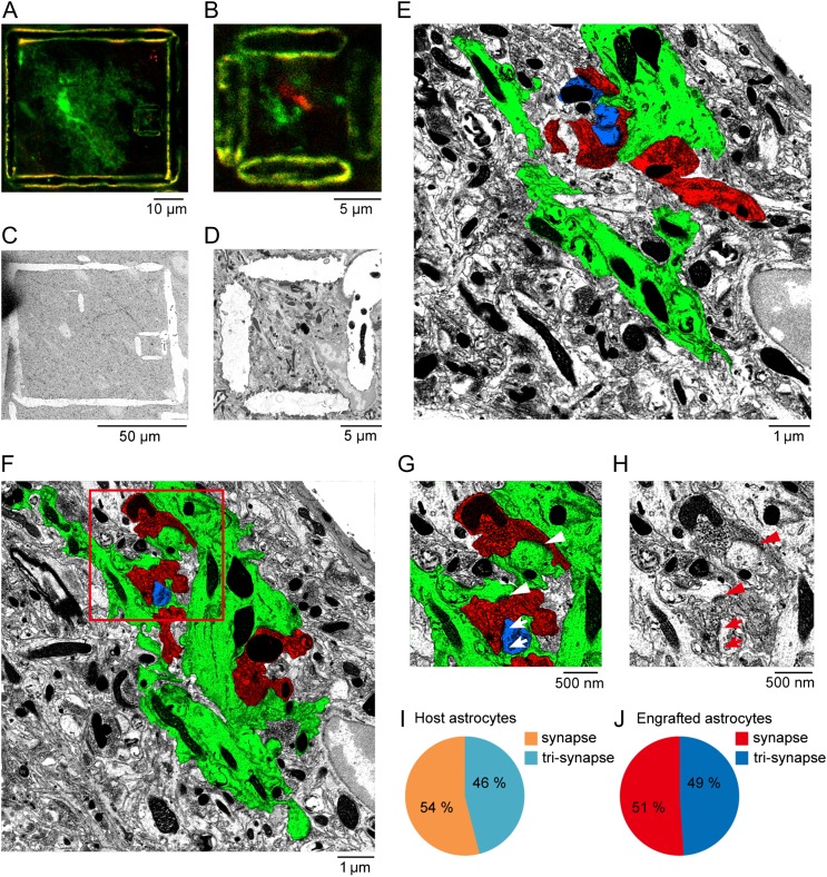
Electron microscopic evidence for the formation of synapse-like contacts between engrafted astrocytes and cholinergic axonal terminals. (A) Two-photon image of EGFP (green) positive engrafted astrocytes and mini-Ruby (red) positive axons projected from the nucleus basalis of Meynert in a cortical section, after NIRB. The large NIRB box outlines an entire engrafted astrocyte, while the small box outlines a juxtaposed site of the process of the engrafted astrocyte (green) with a mini-Ruby labeled axonal site (red). (B) High magnification of the target region outlined by the small NIRB box in panel A. (C) Low-power electron microscopic image of the target region after switching from thick sectioning to ultrathin sectioning. The NIRB markers can be readily observed. (D) High-power electron micrograph of the small NIRB box, the same as in panel B. (E) Ultrathin section of processes of engrafted astrocytes (green) and mini-Ruby labeled axon (red) outlined by the NIRB mark in panel D. False color image shows the processes of engrafted astrocytes in green, mR-labeled axons in red, and a postsynaptic structure of a host neuron in blue. (F) Another example of electron micrograph. (G) High magnification of the region outlined in F, showing both synaptic-like structures (arrowheads) and tripartite synapses among the processes of astrocyte (green), the mR+ axon (red), and the postsynaptic site (blue) (arrows). (H) The actual EM image, corresponding to G. (I, J) Summary of the percentage of synaptic-like structures (synapse) or tripartite synapses (tri-synapse) formed between host astrocytes and neurons (n = 106 synaptic contacts, 3 cells from 2 mice), or between engrafted astrocytes and neurons (n = 121 synaptic contacts, 3 cells from 2 mice).

