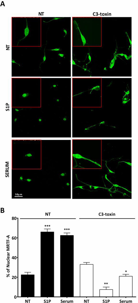
Figure 4. S1P induces MRTF-A accumulation in the nucleus in a Rho dependent manner.
Serum starved mouse CPCs were stimulated with S1P (3µM) or serum (20%) with or without pre-treatment with C3 toxin (1ug/ml). MRTF-A was localized by immunofluorescence; z-stack confocal images were compressed for analysis. The percentage of cells with nuclear MRTF-A was quantified by counting 10 fields in 3 independent experiments (n=50 cells). (A) Representative immunofluorescence of MRTF-A localization Green- MRTF-A (B) Quantification of MRTF-A nuclear accumulation. *p≤0.05 **p≤0.01, and *** p≤0.005 vs. non-treated (NT) by unpaired t-test

