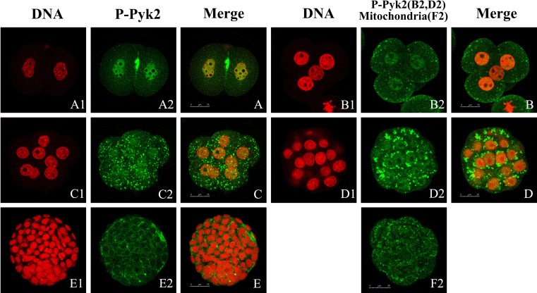
Fig. 4.
Tyr402 phosphorylation Pyk2 during embryonic development. Embryos at the 2- (A1, A2, and A), 4- (B1, B2, and B), and 8-cell (C1, C2, and C) stage; morula (D1, D2, and D); and blastula (E1, E2, and E) were collected and stained using specific antibody against phosphorylated tyrosine402 Pyk2. Primary antibodies were detected via FITC-labeled secondary antibodies. DNA was stained with PI. Laser scanning confocal microscopy was used to locate Pyk2 (green) and DNA (red). Distribution of mitochondria (green) was analyzed by laser scanning confocal microscopy (F2). Scale bar, 25 μm. At least 30 embryos were scanned perstage and representative images are shown.

