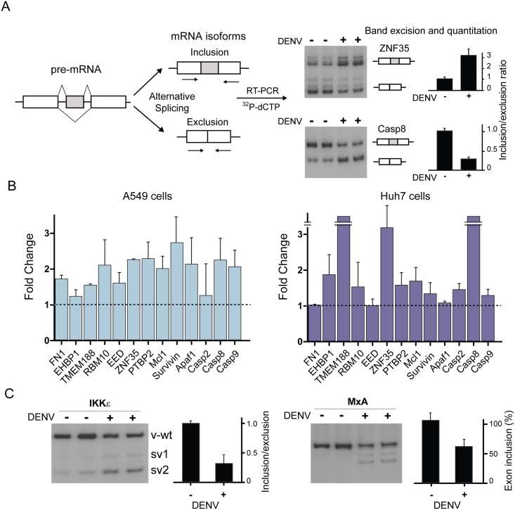
Fig 3. DENV infection modifies the splicing landscape of host cells.
(A) Strategy to detect and quantify alternative splicing isoforms in mock infected or DENV infected cells is depicted on the left. Representative autoradiographs and quantification of the inclusion/exclusion ratio for two alternative exon cassette events are shown (duplicates, mean ± SD). (B) Fold-change of inclusion/exclusion ratios for DENV or mock infected cells. A battery of endogenous alternative exon-cassette events (duplicates, mean ± SD) in A549 and Huh7 cells is shown. (C) DENV-induced changes in the splicing pattern of IKKε and MxA mRNAs. Quantification of alternative splicing events is shown for each case.

