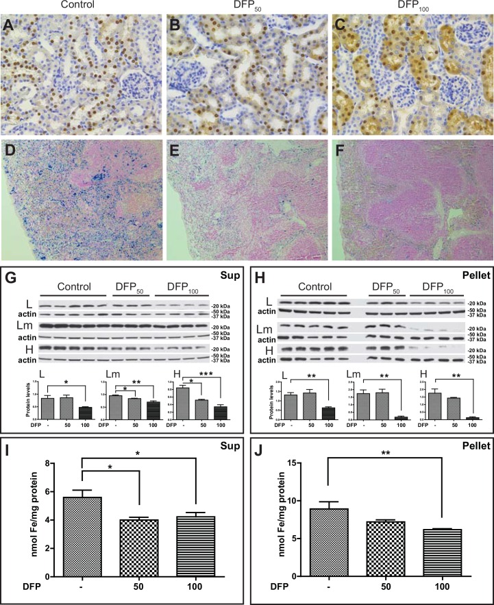
Fig 7. Ferritin and iron deposition in the kidney and spleen of DFP-treated mice.
Histological and immunohistochemical studies of DFP-treated and control FTL-Tg mice (A-F). Analysis of paraffin embedded sections from control (A, D), DFP50 (B, E), and DFP100 (C, F) treated FTL-Tg mice. Sections shown are from kidney (A-C) and spleen (D-F). Sections were immunostained with Abs against the mutant L chain (A-C) or stained with the Perls’ Prussian blue method (D-F). Original magnifications x10 (D-F), x40 (A-C). Western blot analysis of protein samples from the kidney using antibodies specific for the L, Lm, and H chains (B). β-actin was used as loading control. Representative blots are shown for five control, three DFP50, and four DFP100 male mice. Densitometric analysis from three independent experiments shows statistical significant differences between the controls and DFP-treated mice in the supernatant (G) and the pellet (H). (*p < 0.05). By the colorimetric ferrozine method, a decrease in the levels of non-heme iron in the kidney of DFP-treated FTL-Tg mice compared with non-treated FTL-Tg controls was observed in the supernatant (DFP50, p = 0.0190; DFP100, p = 0.0299) (I) and the pellet (DFP100, p = 0.0032) (J).

