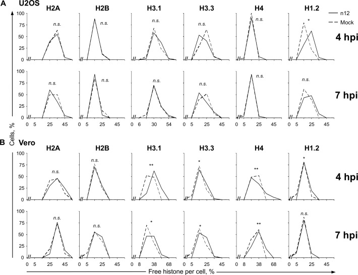
Fig 1. The dynamics of linker and core histones are only minimally altered in the absence of functional ICP4.
U2OS (A) or Vero (B) cells were transfected with plasmids expressing GFP fused to H2A, H2B, H3.1, H3.3, H4, or H1.2. Transfected cells were mock infected or infected with 30 plaque forming units (PFU) per cell of HSV-1 strain n12 and histone dynamics were examined from 4 to 5 or 7 to 8 hours post infection (hpi) (4 hpi or 7 hpi, respectively) by FRAP. Frequency distribution plots showing the percentage of free GFP-H2A, -H2B, -H3.1, -H3.3, -H4, or -H1.2 per individual mock- (dashed line) or n12 (solid line) infected cell at 4 or 7 hpi. **, P < 0.01; *, P < 0.05; n.s., not significant. n ≥ 20 cells from at least 3 independent experiments, except for U2OS H2A (n = 20 cells from 2 independent experiments).

