Fig. 1.
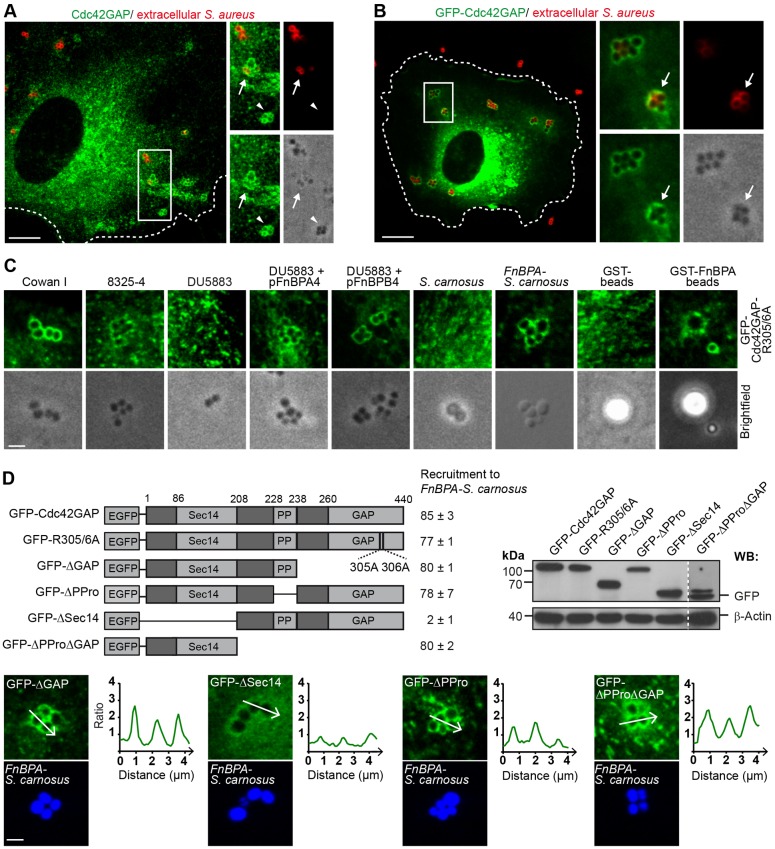
Cdc42GAP is recruited to S. aureus during invasion of HUVECs. (A) HUVECs were infected with S. aureus (Cowan I) for 30 min and immunostained for endogenous Cdc42GAP (green) and extracellularly localized bacteria (red). The boxed region in the overview is depicted as a 1.5-fold enlargement in the right panels [overview (top left), extracellular bacteria (top right), endogenous Cdc42GAP (bottom left) and brightfield (bottom right)]. Cdc42GAP localization at an intracellular S. aureus cluster (white arrowhead) and a partially internalized cluster (white arrow) are highlighted. The cell border is indicated by the broken line. Scale bar: 10 µm. (B) HUVECs expressing GFP–Cdc42GAP were infected with S. aureus (Cowan I) for 30 min and extracellularly localized bacteria were immunostained (red). The boxed region in the overview is depicted as a 2.8-fold enlargement in the right panels [overview (top left), extracellular bacteria (top right), GFP–Cdc42GAP (bottom left) and brightfield (bottom, right)]. GFP–Cdc42GAP localization at a partially internalized cluster is indicated by the white arrow. The cell border is indicated by the broken line. Scale bar: 10 µm. (C) HUVECs expressing GFP–Cdc42GAP-R305/6A were challenged with S. aureus strain Cowan I, S. aureus strain 8325-4, S. aureus strain DU5883, S. aureus strain DU5883 complemented with plasmids pFnBPA4 or pFnBPB4, S. carnosus strain TM300, S. carnosus strain TM300 expressing FnBPA of 8325-4 (FnBPA-S. carnosus), or GST- and GST–FnBPA-coated beads (3-µm diameter) for 30 min. Scale bar: 2.5 µm. (D) Top left, scheme of GFP–Cdc42GAP constructs used in this study: GAP-activity-deficient arginine mutant (GFP-R305/6A); GAP-domain-deficient construct (GFP-ΔGAP); polyproline-region-deficient construct (GFP-ΔPPro); Sec14-domain-deficient construct (GFP-ΔSec14) and polyproline region plus GAP-domain-deficient construct (GFP-ΔPProΔGAP). Recruitment of GFP–Cdc42GAP constructs to FnBPA-S. carnosus is expressed as a percentage of cell-associated bacteria (mean±s.d.). At least 300 bacterial clusters from two different experiments were evaluated per construct. Recruitment was considered positive, when a clear intensity peak of fluorescence adjacent to the bacteria was detected versus background as shown in the intensity plots for GFP–ΔGAP, GFP–ΔPPro and GFP-ΔPProΔGAP in the lower panels. The intensity plot of GFP–ΔSec14 was considered as showing no specific recruitment. Scale bar: 2 µm. Top right, expression of GFP constructs was verified by anti-GFP western blotting (WB). A threefold higher exposure time was used for GFP–ΔPProΔGAP.

