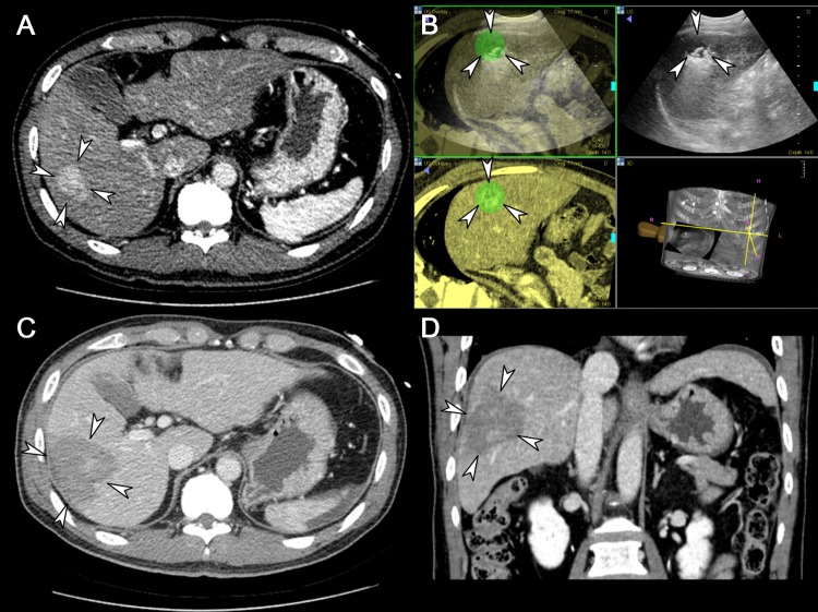
Fig 4. A representative case showing the usefulness of a separable cluster electrode in ablating a large volume at one time.
(A) Axial CT image taken prior to RFA demonstrates a 3.4 cm sized, hypervascular lesion in the right lobe of the liver (arrowheads). (B) Intra-procedural US images fused with pre-procedural CT images guide the tumor (arrowheads) targeting and monitoring. (C) Axial CT image acquired immediately after RFA shows the ablation zone (arrowheads) sufficiently covering the index tumor, measured as 6.0 cm in long diameter, including the safety margin. (D) Coronal CT image reconstructed from the immediate post-procedural CT scan also depicts the ablation zone (arrowheads) measured as 5.9 cm in its coronal long axis.

