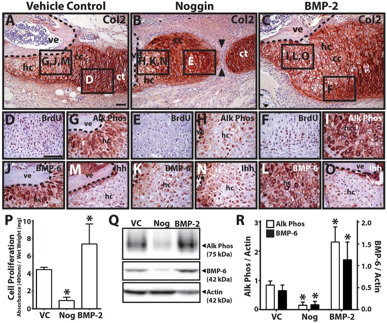
Fig. 6.
BMP inhibition and stimulation modulates proximal cartilage proliferation and hypertrophy. (A-C) Lizard tail blastema explants (9 DPA) were treated with vehicle control (A), noggin (B) or BMP-2 (C) and analyzed by Col2 immunostaining to assess the role of hedgehog signaling in proximal CT hypertrophy. Noggin treatment resulted in non-unions between proximal and distal CT regions (black arrowheads). (D-O) Higher magnification views of regions identified in A-C analyzed by BrdU (proliferating cells; D-F), Alk Phos (G-I), BMP-6 (J-L) and (M-O) Ihh immunostaining. Dashed lines trace vertebral boundaries. (P) Quantification of cell proliferation in CC explants treated with vehicle control (VC), noggin (Nog) or BMP-2 (n=3). *P<0.05 compared with vehicle control; Student's t-test. (Q) Western blot analysis of Alk Phos and BMP-6 expression in CC explants treated with VC, noggin or BMP-2. Actin blots were included as loading controls. (R) Densitometric quantification of Alk Phos and BMP-6 western blot results normalized to actin loading controls (n=3). *P<0.05 compared with vehicle controls; Student's t-test. Data are mean±s.d. cc, cartilage callus; ct, cartilage tube; hc, hypertrophic chondrocytes; ve, vertebra. Scale bars: 50 µm.

