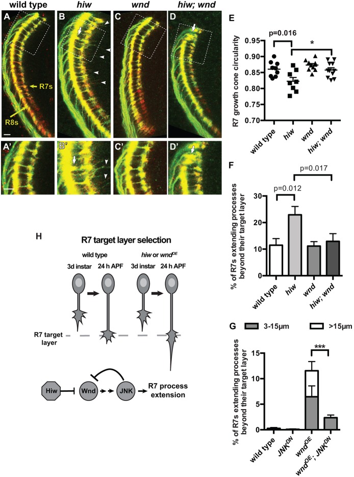
Fig. 2.
R7 growth cones require Hiw to repress Wnd as they halt at their target layer. (A-D′) Pupal medullas (24 h APF; 25°C) in which R7 and R8 axons are labeled with chp-Gal4, UAS-mCD8-GFP (green) and anti-Chp (red). A′-D′ are enlargements of the boxed regions in A-D with enhanced brightness. Yellow arrows in A indicate the R7 and R8 target layers. (E-G) Quantifications of R7 growth cone phenotypes. hiw mutant R7 growth cones are elongated (B,B′,E; two-tailed t-test) and extend processes beyond their target layer (arrowheads; processes of at least 3 µm are quantified in F; two-tailed t-test). R8 growth cones occasionally terminate between the R8 and R7 target layers (arrows in B,B′; 1.7±0.6% versus <0.08% in wild type; Fig. S2B,B′). Loss of wnd has no effect on wild-type R7 growth cones (C,C′,E,F) but restores the morphology of hiw mutant R7 growth cones to that of wild type (D-F; one-tailed t-tests, *P<0.01). R8 growth cones also occasionally extend beyond their target layer in both wnd (data not shown) and hiw; wnd mutants (arrows in D,D′). (G) Using chp-Gal4 to drive co-expression of Wnd (wndOE) and RFP (as a control for UAS copy number) is sufficient to disrupt R7 growth cone halting. Co-expressing Wnd with JNKDN ameliorates this defect (G; ***P<0.0001; Fig. S2). Error bars represent s.e.m. n=9, 8, 11 and 11 brains, from left to right on the graphs in E,F and 7, 8, 7 and 12 from left to right on the graph in G. Scale bars: 5 µm. (H) Model summarizing the roles of Hiw, Wnd and JNK as R7 growth cones halt at their target layer.

