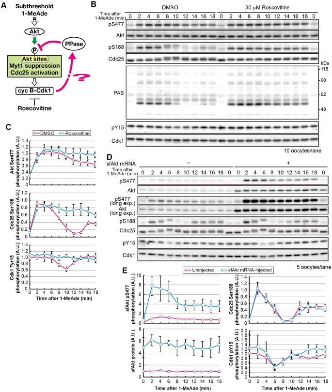
Fig. 2.
The characteristic dephosphorylation of Akt substrates after treatment with subthreshold concentrations of 1-MeAde depends on cyclin-B–Cdk1 activity. (A) Hypothesis tested in B and C. (B) Immature oocytes were treated with subthreshold levels of 1-MeAde (30 nM) in the presence of 30 μM roscovitine or 0.15% DMSO (as a negative control) and collected at the indicated times, followed by immunoblotting to detect phosphorylation and total protein levels. (C) Phosphorylation levels were quantified from the images in B, as described in Fig. 1C. Data represent mean values±s.d. from three independent experiments. (D) Immature oocytes were injected with mRNA encoding starfish Akt (sfAkt), incubated for 3 h, treated with a subthreshold concentration (30 nM) of 1-MeAde, and collected at the indicated times, followed by immunoblotting for phosphorylation and total proteins. (E) Phosphorylation levels were quantified from the images in D and normalized against the total amounts of each protein. Phosphorylation levels relative to those in uninjected oocytes at 2 min are shown. Data represent mean values±s.d. from three independent experiments. cyc, cyclin; exp., exposure; PPase, phosphatase.

