
Fig. 3.
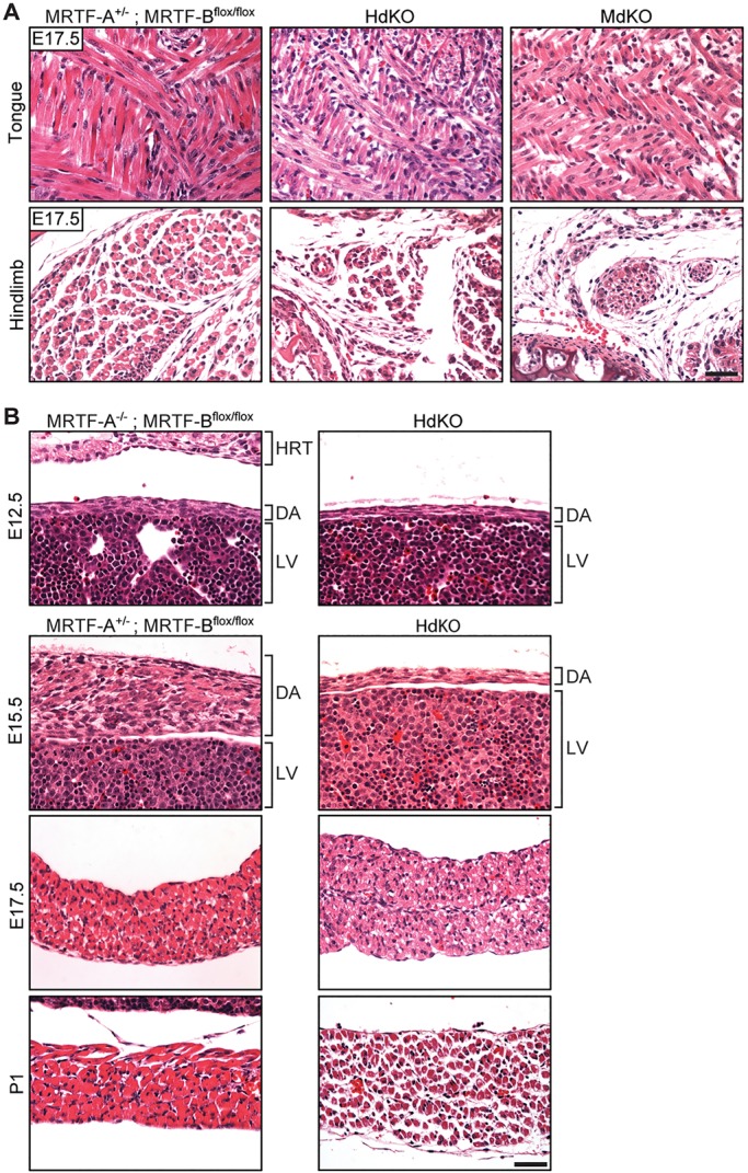
Hypoplasia of myofibers is observed in embryonic muscle tissue of MRTF dKO animals. (A) H&E staining of coronal sections of tongue and transverse sections of hindlimb from E17.5 control (MRTF-A+/−; MRTF-Bflox/flox) and dKO embryos shows a reduction in myofiber size in the dKO tissue. (B) H&E staining of WT and MRTF dKO diaphragm at E12.5, E15.5, E17.5 and P1 shows myofiber hypotrophy at all ages that becomes more pronounced with age. DA, diaphragm; HRT, heart; LV, liver. Scale bars: 40 μm.