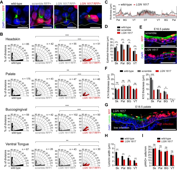
Fig. 3.
Altered spindle orientation and differentiation upon LGN loss. (A) Representative wild-type littermate, scramble, LGN 1617 RFP− and LGN 1617 RFP+ telophase palatal cells indicating division orientation (yellow angle) relative to the BM (dashed line). (B) Radial histograms of division orientation for headskin, palatal epithelium, BG epithelium and VT at E16.5. Perpendicular and planar divisions are roughly equal in wild-type, LGN 1617 RFP− and scramble cells, whereas oblique and planar divisions are increased in LGN 1617 RFP+ mitoses. (C) Average OE thickness in wild-type and LGN knockdown embryos at E16.5, measured every 10 μm and averaged from three embryos per group. (D) Epithelial thickness for the indicated tissues in control groups (wild-type littermates and scramble) and LGN knockdowns (red). (E,F) Reduced immunolabeling for the spinous/granular marker K10 (green in E) in E16.5 LGN knockdowns, as quantified in F. Basal (K14) marker expression is unaffected. (G) Differentiation defects at E18.5 showing reduced loricrin immunolabeling specifically in RFP+ regions of LGN knockdown palatal epithelium. (H) Similar reductions in loricrin staining are observed across all OE. (I) LGN loss does not significantly impact proliferation rates. (D,F,H,I) Data are derived from n=10-30 sections per tissue per age from n≥4 independent embryos. *P<0.05, **P<0.01, ***P<0.0001, by χ2 test (B) or Student's t-test (D,F,H,I); ns, not significant. Scale bars: 10 µm in A; 50 µm in E,G.

