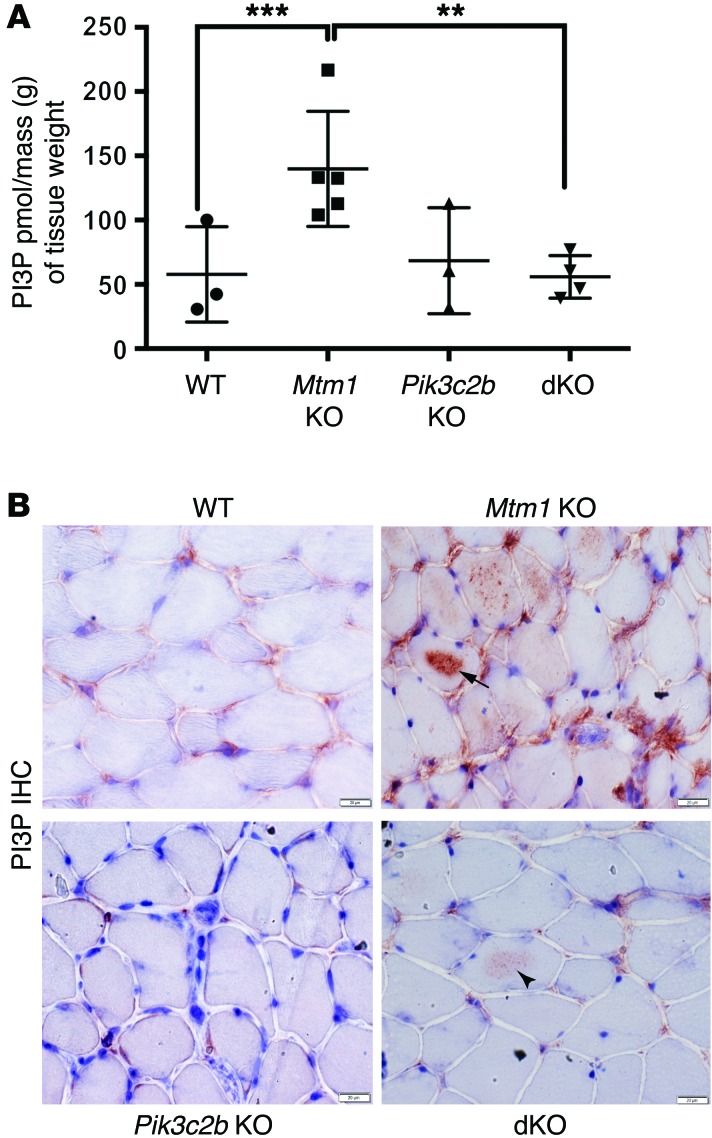
Figure 3. Restoration of PI3P levels in the Pik3c2b Mtm1 dKO mice.
(A) PI3P levels are normalized in the dKO animals, as determined using a PI3P ELISA kit [purified lipid (pmol)/mass (g) of muscle tissue]. PI3P levels in WT muscle = 58 ± 21 pmol/g (n = 3), Mtm1 KO = 140 ± 20 pmol/g (n = 5, ***P = 0.01 vs. WT, **P = 0.01 vs. dKO), Pik3c2b KO = 68 ± 24 pmol/g (n = 3), and dKO = 56 ± 8 pmol/g (n = 4, P = 0.87 vs. WT, 1-way ANOVA followed by Dunnett’s multiple comparison test). (B) PI3P subcellular localization by IHC on tibialis anterior muscle. Labeling in WT and Pik3c2b KO is observed faintly in the perinuclear and membrane compartment, while staining in Mtm1 KO is seen diffusely, with intense intracellular staining in the sarcolemma of several myofibers (arrow). dKO muscle shows staining similar to WT, though with areas of mildly increased labeling within the sarcolemma of selected myofibers (arrowhead). Scale bars: 20 μm.

