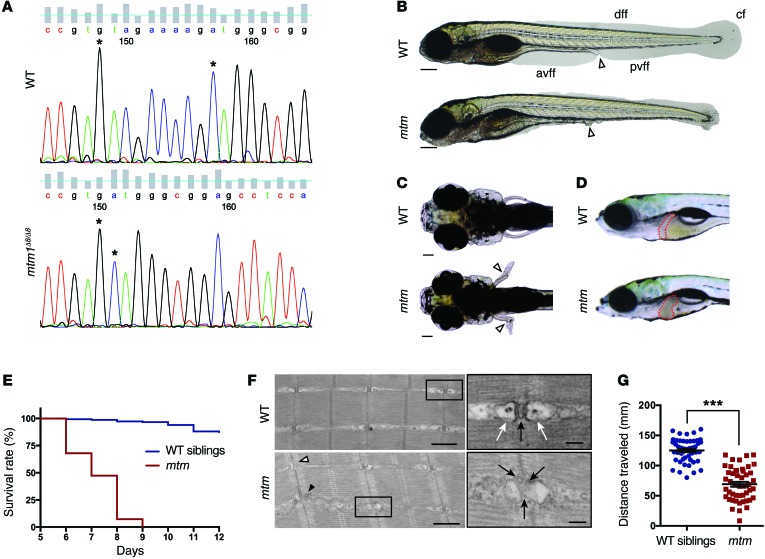
Figure 6. Phenotypic characterization of mtm, a novel zebrafish model of MTM.
(A) Sanger sequencing of an RT-PCR fragment from WT (top) and homozygous mtm1Δ8/Δ8 mutant (mtm; bottom) larvae showing an 8-bp deletion in exon 5 of mtm1. (B) Bright-field of WT siblings compared with mtm larvae at 4 dpf illustrating complete loss of the anterior and posterior ventral fin folds (white arrowhead indicates urogenital opening; scale bar: 200 μm). This represents a “severe” mutant phenotype. avff, anterior ventral fin fold; pvff, posterior ventral fin fold; dff, dorsal fin fold; cf, caudal fin. (C and D) By 7 dpf, mtm larvae have outwardly kinked pectoral fins (white arrowheads; scale bar: 100 μm) and abnormal liver appearance (liver outlined in red). (E) mtm larvae have median and maximum survival of 7 and 9 days, respectively, while most WT siblings survive into adulthood (n = 150 per group; P < 0.001, Mantel-Cox test). (F) At 5 dpf, electron micrographs show aberrant skeletal muscle ultrastructure in mtm larvae (scale bars: 500 nm; inset: 100 nm). WT larvae have normal triad structure where T-tubules (inset; black arrow) are apposed by terminal cisternae/SR (inset; white arrows), whereas mtm muscle has L-tubules (black arrowhead), triads lacking SR (white arrowhead), and fragmented T-tubules (inset; black arrows). (G) mtm larvae have significantly impaired motor function compared with WT. In a 30-second optovin-induced photoactivation period performed at 7 dpf, mutants only travel about half (55%) the distance covered by their siblings (***P < 0.001). n = 48 each; Student’s t test, 2-tailed. Error bars indicate SEM.

