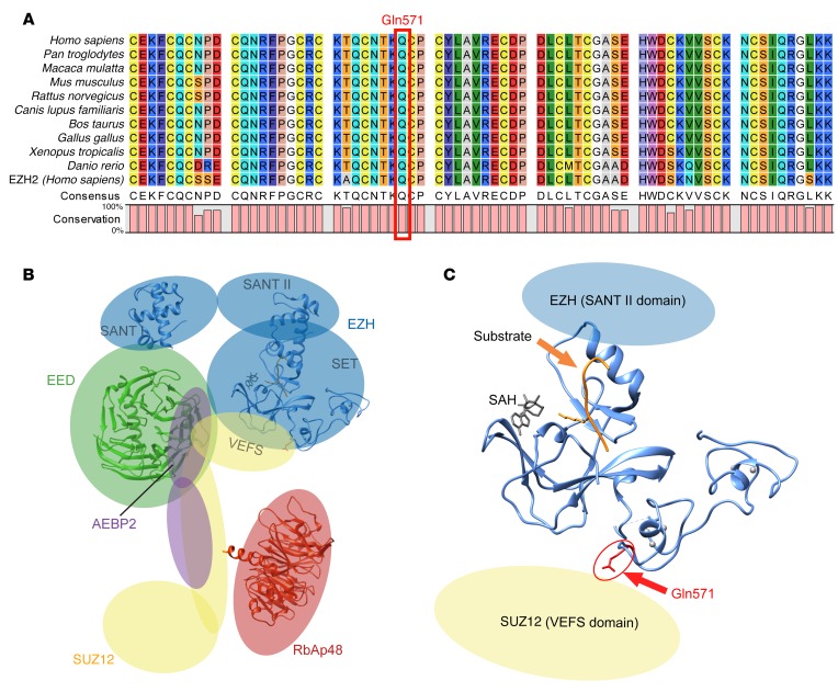
Figure 2. Location of the Gln571Arg mutation in EZH1.
(A) Sequence alignment of the region corresponding to Gln571 in EZH1 from different species. The sequence of the closely related human EZH2 is included. (B) Predicted overall structure of PRC2 (based on ref. 18). (C) Position of the residue corresponding to Gln571. Since no structural data are available for EZH1, a structure of the catalytic domain of EZH2 (Protein Data Bank [PDB] ID: 4MI5; 95% homology) is shown. The positions of the substrate and cofactor S-adenosyl-L-homocysteine (SAH) are based on an alignment with human H3K9 methyltransferase (PDB ID: 3HNA).

