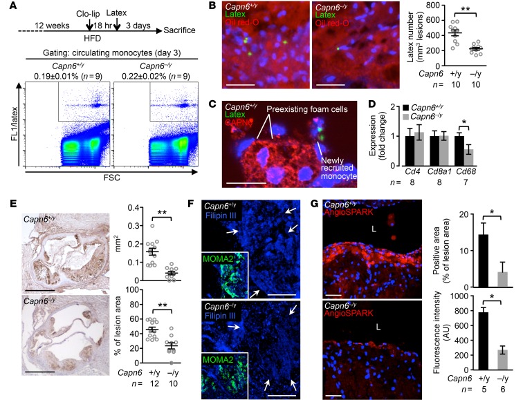
Figure 10. Ablation of CAPN6 diminishes the recruitment of macrophages and their pinocytotic ability in murine atherosclerotic lesions.
(A) Uptake of latex beads in circulating monocytes at day 3 was equivalent between Capn6+/yLdlr–/– and Capn6–/yLdlr–/– mice. (B) Latex-positive monocytes in aortic atherosclerotic lesions were reduced by Capn6 deficiency. (C) CAPN6 expression is abundant in preexisting foam cell macrophages, but not in newly recruited macrophages, in Capn6+/yLdlr–/– atheromas. (D) PCR-based quantification of leukocyte markers in aortic atherosclerotic lesions in mice receiving HFD for 12 weeks. (E) Deposition of macrophages in atherosclerotic lesions is reduced by Capn6 deficiency. MOMA2+ area in aortic sinus lesions in the mice receiving HFD for 12 weeks was quantified. (F) Cholesterol deposition in atherosclerotic plaques. Aortic sections were stained with Filipin III. Arrows indicate macrophage-enriched regions. (G) Pinocytotic activity in atherosclerotic lesions. AngioSPARK nanoparticles were i.v. injected as a pinocytotic activity marker. L, aortic lumen. **P < 0.01; *P < 0.05; Student’s t test (D, E, and G) and Mann-Whitney U test (B); error bars represent mean ± SEM. Scale bars: 50 μm (B); 20 μm (C); 500 μm (E); 100 μm (F); 40 μm (G).

