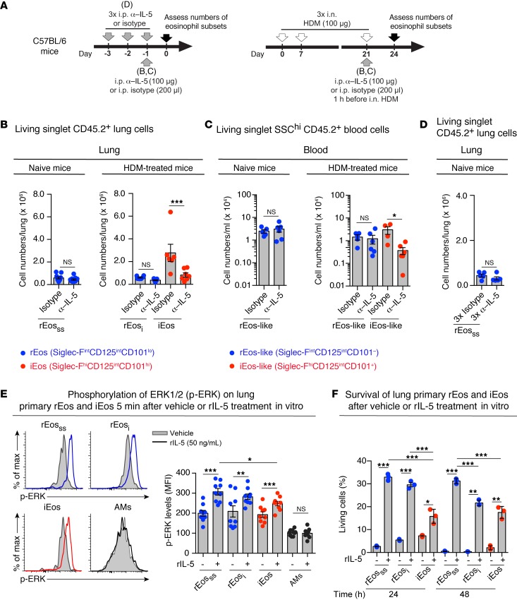
Figure 6. Sensitivity and responsiveness of eosinophil subsets to in vivo α–IL-5 and in vitro rIL-5 treatments.
(A) Experimental outline for data shown in B–D. (B–D) Effects of in vivo α–IL-5 (TRFK5) treatment on the numbers of (B and D) lung and (C) blood eosinophil subsets in naive and HDM-treated allergic C57BL/6 mice. (E) Phosphorylation of ERK1/2 was assessed by phospho-flow cytometry on freshly isolated eosinophils and AMs stimulated in vitro with rIL-5 or vehicle. Representative flow cytometric histograms and quantification of p-ERK levels. (F) Survival (as assessed by incorporation of 7-AAD) of FACS-sorted lung eosinophil subsets stimulated ex vivo, with or without rIL-5, for the indicated times. More than 90% of the cells were negative for 7-AAD before stimulation. (B–F) Data represent the mean ± SEM as well as individual values and are pooled from 2 to 3 independent experiments (n = 5–11/group). *P < 0.05, **P < 0.01, and ***P < 0.001, by 2-tailed Student’s t test (B and C, left panels, and D) or a mixed-effects model, followed by Tukey’s test for multiple comparisons (B and C, right panels) on log-transformed values and (E and F) by Welch’s t test (for comparison of vehicle vs. IL-5–treated groups) or 1-way ANOVA, followed by Tukey’s test for multiple comparisons (for comparison of IL-5–treated subsets). cpm, counts per minute.

