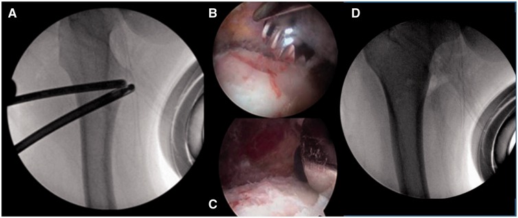
Fig. 4.
(A–D) Fluoroscopic view of the right hip showing the 5.5 mm burr placed on the anterior surface of the lesser trochanter; arthroscopic views (B–C) showing the progressive resection of the lesser trochanter; and a fluoroscopic view (D) demonstrating the completed resection of the lesser trochanter.

