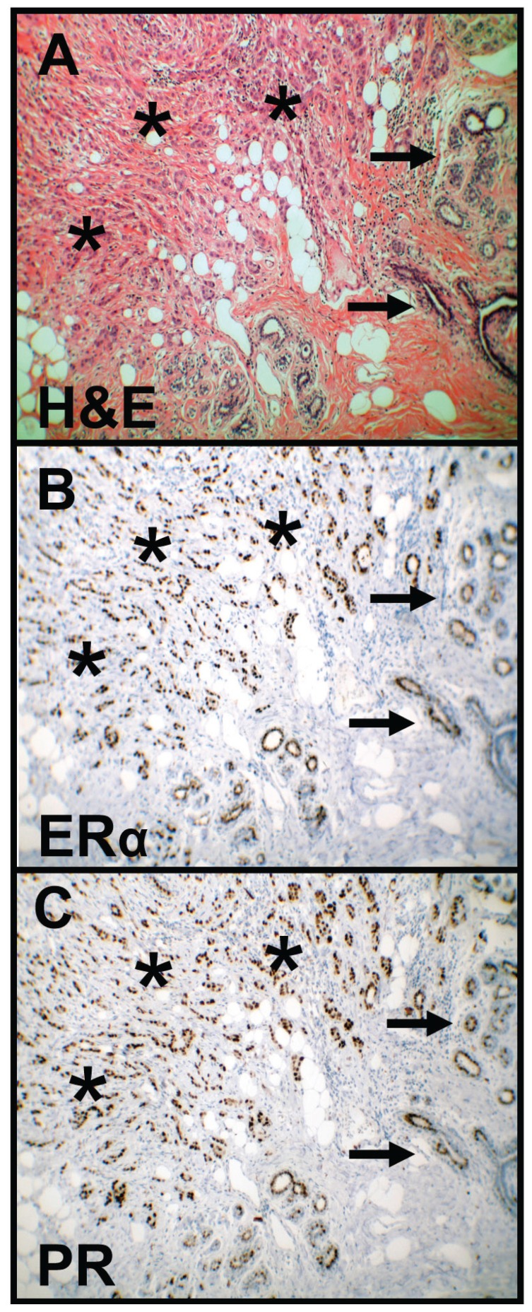
Figure 1.
HE staining and immunohistochemical staining of ERα and PR in breast cancer. HE staining (A) shows tumor cells (asterisks) and normal mammary gland tissue (arrows). Immunohistochemical staining shows representative images of positive ERα (B) and PR (C) expression in breast cancer as a positive control (asterisks). Brown chromogen color (3,3’-Diaminobenzidine) indicates positive nuclear staining, the blue color shows the nuclear counterstaining by hematoxylin (original magnification: x100-fold). HE, Haematoxylin and eosin; ERα, Estrogen Receptor alpha; PR, Progesterone Receptor.

